

Abstract
Total pelvic exenteration with extended endo-pelvic resection is an extensive oncologic procedure associated with prolonged operative duration and major blood loss. Perioperative management becomes more complex when the tumor involves large vessels. We report the perioperative management of a 54-year-old hypertensive woman with recurrent uterine sarcoma (low grade stromal sarcoma) who underwent the procedure. The tumor encased the right external iliac, internal iliac, and common iliac veins, with thrombus extending to the bifurcation of the inferior vena cava. Intraoperative blood loss was approximately 6.7–7 liters. She received 11 units packed red blood cells (PRBC), 10 units fresh frozen plasma (FFP), 6 unit’s random donor platelets, 8 units cryoprecipitate, 10 unit’s crystalloids and one unit of 20% human albumin with operative duration of 11hours. A 12 Fr Mahurkar dialysis catheter was placed in the right internal jugular vein to facilitate rapid transfusion. The case highlights pelvic vascular anatomy, massive transfusion protocol, vascular access strategy, electrolyte management, long duration total intravenous anesthesia, analgesic planning and airway strategy. Coordinated teamwork was central to management.
Keywords
Pelvic exenteration, Uterine sarcoma, Dialysis catheter, Peri-operative management, Massive blood transfusion, Coagulopathy, Team work
Abbreviations
TPE: Total Pelvic Exenteration; TIVA: Total Intravenous Anesthesia; MTP: Massive Transfusion Protocol; IVC: Inferior Vena Cava; PRBC: Packed Red Blood Cells; FFP: Fresh Frozen Plasma; TAH+BSOT: Total Abdominal Hysterectomy Bilateral Salphingoophorectomy; SUVmax: Standardized Uptake Value Maximum; FDG: Fluorodeoxyglucose; INR: International Normalized Ratio; APTT: Activated Partial Thromboplastin Time; TEG: Thromboelastography; ROETM: Rotational Thromboelastometry; NSAID: Non-Steroidal Anti-Inflammatory Drug; ICU: Intensive Care Unit; ABG: Arterial Blood Gas; BUN: Blood Urea Nitrogen; PET-CT: Positron Emission Tomography Computed Tomography; BMI: Body Mass Index; BSA: Body Surface Area
Introduction
Total pelvic exenteration (TPE) is performed for locally advanced or recurrent pelvic malignancies when disease remains confined to the pelvis but is not amenable to limited resection [1]. The procedure involves En bloc removal of pelvic organs and extensive dissection near major vascular structures, often resulting in significant blood loss and prolonged anesthesia. Uterine sarcomas are rare mesenchymal tumors characterized by aggressive behavior, early recurrence, and a tendency for local invasion. Unlike endometrial carcinoma, recurrence commonly occurs within the pelvis with infiltration of adjacent organs and vascular structures [2]. Following hysterectomy, therapeutic options for recurrent disease are limited, and exenterative surgery may be considered in carefully selected patients. We report on the peri-operative anesthetic management of a patient with recurrent uterine sarcoma with major vascular encasement undergoing TPE.
Case Presentation
A 54-year-old woman with Body mass index (BMI) of 20.3 kg/m2 and Body surface area (BSA) 1.52 m2 presented with recurrent pelvic malignancy (low grade stromal sarcoma). She was a known hypertensive on regular medication. She had undergone total abdominal hysterectomy with bilateral salpingo-oophorectomy for uterine sarcoma. Subsequently, she required partial cystectomy. She presented with abdominal distension and bleeding per-vagina. She had undergone radiation, chemotherapy (carboplatin, paclitaxel) and hormonal therapy post Total Abdominal Hysterectomy Bilateral Salphingoophorectomy (TAH+BSO). Preoperative Positron Emission Tomography Computed Tomography (PET–CT) demonstrated a large heterogeneous solid-cystic pelvic mass with mild FDG avidity Standardized Uptake Value maximum (SUVmax ~5.7), extensively infiltrating adjacent pelvic structures including the rectum, sigmoid colon, pelvic sidewall, pelvic floor muscles, urethra, and vaginal vault. The lesion partially encased the right iliac vessels with tumor thrombus extending from the right external and common iliac veins up to the inferior vena cava bifurcation. Superior extension involved the right psoas muscle and sacral nerve roots. Bilateral distal ureteric infiltration caused hydro-ureteronephrosis, with fistulous communications between the ureter and sigmoid colon as well as a pelvic collection. Additional findings included Fluorodeoxyglucose (FDG) avid retroperitoneal and inguinal lymph nodes, transverse loop colostomy in situ, and bilateral percutaneous nephrostomy tubes. There was no evidence of distant metastasis. Total pelvic exenteration was planned.
Diagnostic Assessment
The patient was hemodynamically stable preoperatively. Baseline hemoglobin of 10.9 g/dl, platelet count of 2,16,000cells/µL and coagulation profile (PT- 12.2 test, 12.2 control, INR- 1.3, aPTT- 32) were noted. Renal function was preserved with functioning nephrostomies and electrolytes were normal (Sodium-132, Potassium-3.4, Calcium-8.6). Preoperative cardiorespiratory evaluation, including ECG and 2D echocardiography, showed no significant abnormalities except well-controlled hypertension. Preoperative hemoglobin of 10.9 g/dL was considered acceptable and did not require further optimization. The patient had already received iron supplementation and prior transfusions, with no symptoms attributable to anemia. Given the urgency of surgery and advanced malignancy, additional measures such as erythropoietin were unlikely to provide significant benefit. Hemoglobin levels of 10–12 g/dL are also commonly encountered in the Indian population and were considered adequate for proceeding with surgery [3,4]. The presence of major venous encasement and Inferior Vena Cava (IVC) thrombus raised concerns regarding hemorrhage, hemodynamic instability and possible thromboembolic events. Anticipated duration of surgery was prolonged. A multidisciplinary discussion was conducted involving surgical oncology, vascular surgery, anesthesia, intensive care, and transfusion services. Blood products were arranged in advance, and a massive transfusion protocol was kept ready for activation.
Anesthetic Plan
Oral intubation with controlled mechanical ventilation under Total intravenous anesthesia (TIVA) with all standard ASA monitors and invasive hemodynamic monitoring.
Therapeutic Intervention
On the day of surgery, standard monitoring was instituted and no overnight fluid at maintenance rate was administered. A radial arterial catheter was inserted for continuous blood pressure monitoring and serial blood sampling. Considering the likelihood of rapid blood loss, (maximum allowable blood loss calculated-291 ml, weight-52 kg, trigger Hb-10 g/dl) vascular access was prioritized before incision. Instead of a conventional triple lumen central venous catheter, a 12 Fr Mahurkar dialysis catheter was inserted into the right internal jugular vein under ultrasound guidance [5]. The placement of a 12 Fr Mahurkar dialysis catheter was a key component of the anesthetic strategy, providing robust high-flow vascular access that enables rapid large-volume transfusion during catastrophic blood loss and plays a critical role in maintaining hemodynamic stability throughout the procedure. Additional large-bore peripheral intravenous cannulas were secured. General anesthesia was induced with intravenous propofol and opioid, followed by neuromuscular blockade to facilitate endotracheal intubation. Epidural was placed at T10–11ideally considering mid-line laparotomy incision [6]. The catheter was placed before the anticipated hemorrhagic phase of surgery and was intended to provide perioperative and postoperative analgesia. Following the onset of massive hemorrhage and coagulopathy, epidural management adhered to neuraxial safety guidelines, and catheter removal was deferred until coagulation abnormalities had resolved. Although the presence of an IVC-level thrombus increased surgical complexity, it did not represent an absolute contraindication to neuraxial anesthesia [7]. The decision was made after multidisciplinary discussion involving anesthesiology and the surgical team, balancing the anticipated analgesic benefits against the potential risks. Transesophageal echocardiography (TEE) and other cardiac output monitoring modalities were not available intraoperatively. Anesthesia was maintained using TIVA with propofol infusion combined with opioid infusion [8]. This technique was selected to provide hemodynamic control during prolonged surgery and to allow predictable recovery, supported by tranexamic acid bolus of 1 gm followed by infusion at 1 mg/kg/hr. Neuromuscular blockade was maintained as required. Active warming measures were instituted using a forced-air warming system and fluid warmers to reduce risk of hypothermia. Temperature, urine output, and serial arterial blood gases were monitored throughout.
Intraoperative Course and Massive Transfusion
Surgical dissection was prolonged and technically demanding due to prior surgeries and tumor infiltration. During vascular dissection, significant venous bleeding occurred 4 hours into surgery and was progressively worsening as expected from the pelvic bed. IVC filter placement was avoided as it would have interfered with infrarenal IVC control and because the lesion represented tumor thrombus extending from the external and common iliac veins to the IVC confluence, with a low risk of embolization. The involved venous segment was resected En bloc after obtaining infrarenal IVC control, and primary repair was performed at the right iliac vein–IVC confluence. Reconstruction was not undertaken due to chronic venous occlusion, well-developed collateral circulation, and the high risk of graft thrombosis. Estimated blood loss reached approximately 6.7–7 liters. Massive transfusion protocol was activated early in the course of hemorrhage [9]. The patient received 11 units of PRBC, 10 units of FFP, 6 units of random donor platelets, and 8 units of cryoprecipitate. Approximately 10 units of crystalloid solutions were administered along with one unit of 20% human albumin. Balanced resuscitation was aimed at maintaining an approximate 1:1:1 ratio between red cells, plasma and platelets to reduce dilutional coagulopathy. Cryoprecipitate was given to maintain fibrinogen levels within acceptable limits. Viscoelastic coagulation monitoring using Thromboelastography, Rotational Thromboelastometry (TEG/ROTEM) and intraoperative cell salvage were not available at our institution; therefore, transfusion and coagulation management were guided by conventional laboratory parameters and clinical assessment. Serial arterial blood gases were used to assess hemoglobin, lactate, base deficit and electrolytes. Sugars were less than 200 mg/dl and didn’t require insulin infusion to counteract stress hyperglycemia. Coagulation parameters were checked intermittently to guide component therapy. Urine output during the procedure was approximately 2 liters, indicating maintained renal perfusion despite significant blood loss. Hemodynamic stability was achieved with volume replacement and intermittent vasopressor Nor-adrenaline dose ranging from 0.025-0.5 mcg/kg/min as required till the end of procedure. Serial ABGs are enclosed in Table 1.
|
Parameter |
1 |
2 |
3 |
4 |
5 |
6 |
7 |
|
pH |
7.240 |
7.249 |
7.434 |
7.432 |
7.328 |
7.429 |
7.455 |
|
pCO2 (mmHg) |
37.7 |
36.8 |
33.3 |
36.4 |
25.9 |
34.0 |
34.1 |
|
pO2 (mmHg) |
268.1 |
250.0 |
257.3 |
235.5 |
49.4 |
168.0 |
211.0 |
|
HCO3- (mmol/L) |
16.1 |
16.1 |
22.3 |
24.3 |
13.6 |
22.0 |
23.4 |
|
BE (ecf) |
-11.2 |
-11.1 |
-2.0 |
0.0 |
-12.4 |
-2.3 |
-0.5 |
|
sO2 (%) |
99.8 |
99.8 |
99.9 |
99.8 |
82.7 |
98.6 |
98.9 |
|
Na+ (mmol/L) |
138 |
137 |
141 |
142 |
134 |
145.0 |
145.1 |
|
K+ (mmol/L) |
3.6 |
4.0 |
3.4 |
3.2 |
3.8 |
3.14 |
3.60 |
|
Ca²+ (mmol/L) |
1.35 |
1.29 |
1.10 |
1.08 |
1.47 |
1.08 |
1.11 |
|
Cl- (mmol/L) |
115 |
114 |
106 |
105 |
110 |
106 |
103 |
|
TCO2 (mmol/L) |
15.7 |
15.7 |
21.3 |
23.2 |
15.1 |
21 |
23 |
|
Anion Gap |
8 |
8 |
15 |
15 |
12 |
20.1 |
22.3 |
|
Anion Gap (K) |
12 |
12 |
18 |
18 |
16 |
18 |
18 |
|
Hct (%) |
45 |
35 |
17 |
25 |
25 |
31 |
39 |
|
cHgb (g/dL) |
15.3 |
11.9 |
5.7 |
8.7 |
8.6 |
10.6 |
10.1 |
|
BE(b) |
-10.5 |
-10.3 |
-1.8 |
0.1 |
-12.4 |
-2.3 |
-0.5 |
|
Glucose (mg/dL) |
148 |
194 |
202 |
146 |
254 |
177 |
174 |
|
Lactate (mmol/L) |
1.81 |
3.30 |
3.83 |
4.03 |
3.11 |
3.99 |
2.57 |
|
BUN (mg/dL) |
13 |
13 |
11 |
12 |
12 |
11 |
12 |
|
Urea (mmol/L) |
4.8 |
4.8 |
4.0 |
4.3 |
4.5 |
4.0 |
4.3 |
|
Creatinine (mg/dL) |
0.64 |
0.69 |
0.64 |
0.62 |
0.68 |
0.64 |
0.64 |
|
Parameter |
Baseline |
Value During Hemorrhage |
End of Surgery |
Interpretation |
|
Duration of surgery |
– |
– |
11 h |
Prolonged major surgery |
|
Estimated blood loss |
0 L |
6.7–7.0 L |
6.7–7.0 L |
Massive hemorrhage |
|
Hemoglobin (g/dL) |
15.3 |
5.7 |
10.1 |
Severe acute blood loss anemia |
|
Hematocrit (%) |
45 |
17 |
39 |
Significant hemodilution/blood loss |
|
Lactate (mmol/L) |
1.81 |
4.03 |
2.57 |
Tissue hypoperfusion, improved after resuscitation |
|
pH |
7.24 |
7.24 |
7.46 |
Metabolic acidosis corrected |
|
HCO3- (mmol/L) |
16.1 |
13.6 |
23.4 |
Improvement in metabolic status |
|
Base Excess (mmol/L) |
-11.2 |
-12.4 |
-0.5 |
Resolution of metabolic acidosis |
|
Noradrenaline (µg/kg/min) |
0.025 |
0.5 |
0.05 |
Escalation during shock, weaned after hemostasis |
|
International normalized ratio (INR) |
1.0 |
2.4 |
1.1 |
Coagulopathy corrected |
|
Activated partial thromboplastin time (aPTT) |
32 |
62 |
30 |
Coagulopathy corrected |
|
Fibrinogen (mg/dL) |
410 |
110 |
260 |
Consumption coagulopathy improved |
|
Platelets (×10³/µL) |
220 |
80 |
160 |
Recovery following transfusion |
|
Vascular Access Device |
Size |
Approximate Flow Rate (mL/min) |
Clinical Use |
|
18G Peripheral Cannula |
18G |
103 |
Routine fluid administration |
|
16G Peripheral Cannula |
16G |
236 |
Moderate-volume resuscitation |
|
14G Peripheral Cannula |
14G |
270 |
Preferred peripheral access for massive transfusion |
|
Triple-Lumen CVC |
7 Fr |
Distal- 35–65 Medial- 15–30 Proximal- 15–30 |
Central access, vasoactive infusions |
|
Dialysis Catheter |
11.5–14 Fr |
300–400ml |
High-flow blood and fluid administration during massive hemorrhage |
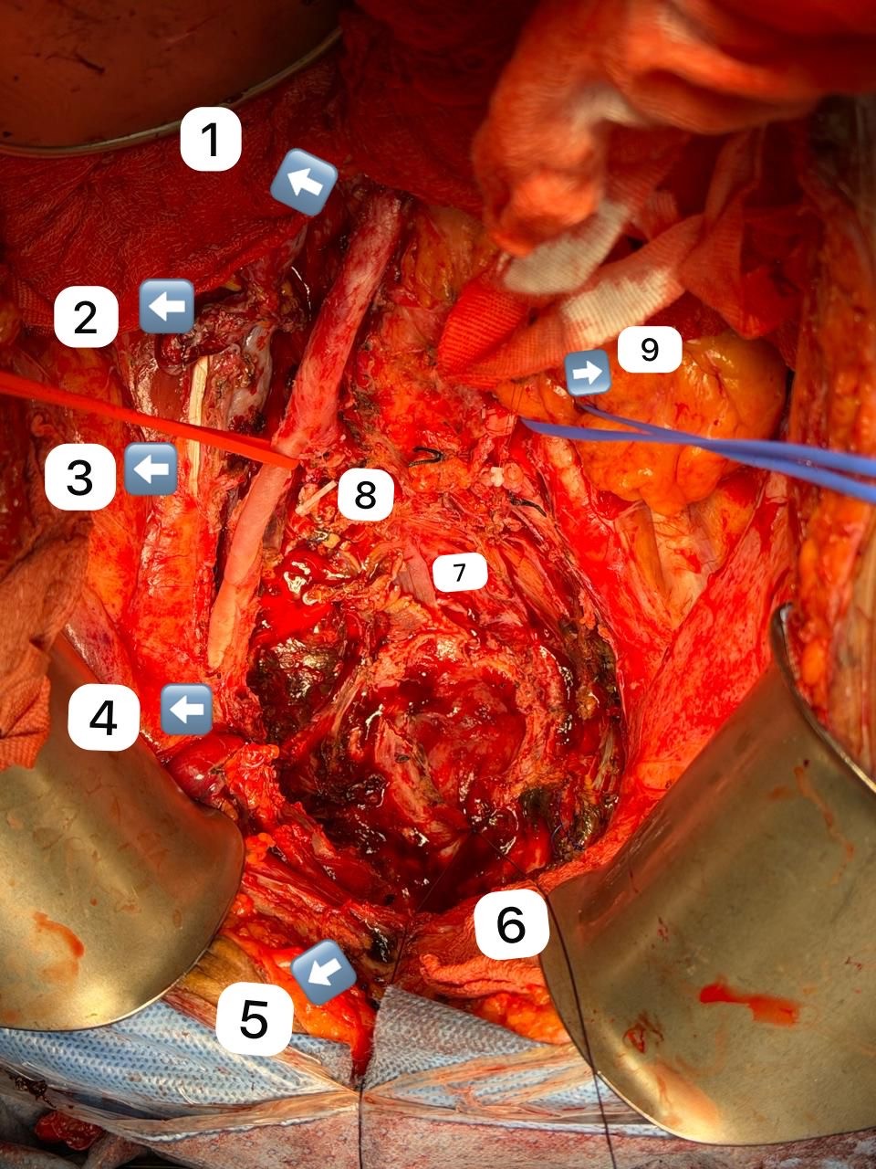
Figure 1.1. Right Common Iliac Artery 2. Psoas Muscle 3. Femoral Nerve 4. Right External Iliac Artery 5. Pubic symphysis 6. Anal Canal margin 7. Sacral nerve roots 8. Ligated Right Internal iliac artery 9. Left common Iliac artery.
Electrolyte and Metabolic Management
Massive transfusion during prolonged oncologic surgery can result in significant metabolic and electrolyte disturbances. Citrate contained in transfused blood products may chelate ionized calcium, predisposing the patient to hypocalcemia, which can impair myocardial contractility and coagulation. Therefore, ionized calcium levels were closely monitored with serial arterial blood gas analyses, and intravenous calcium gluconate supplementation was administered at 30 mg/kg, slow bolus as in when levels declined. Hyperkalemia is another recognized risk due to potassium leakage from stored red blood cells, particularly when large volumes are transfused rapidly. Serum potassium levels were assessed frequently through point-of-care blood gas analysis and remained within acceptable limits throughout the procedure. Metabolic acidosis may occur secondary to tissue hypo perfusion, lactate accumulation, and the metabolic burden of large-volume transfusion. In this case, correction primarily focused on restoration of adequate circulating volume and tissue perfusion through timely blood product replacement and hemodynamic optimization. Sodium bicarbonate was administered selectively when metabolic parameters indicated the need for additional buffering. A key perioperative objective was to prevent the development of the “lethal triad” of hypothermia, acidosis, and coagulopathy that commonly complicates massive hemorrhage. Active warming strategies were used to maintain normothermia, and transfusion therapy was guided by laboratory parameters to reduce dilutional coagulopathy. Continuous metabolic monitoring and prompt correction of abnormalities were essential to maintaining physiologic stability during this prolonged high-risk procedure [10].
Long Duration Anesthesia Considerations
Total pelvic exenteration is a prolonged and physiologically demanding procedure that may continue for many hours. Extended periods of anesthesia increase the likelihood of complications such as pressure injuries, peripheral nerve compression, hypothermia, and disturbances in fluid and electrolyte balance. Consequently, careful intraoperative planning and vigilant monitoring are necessary to reduce these risks. Positioning was performed with particular attention to protecting vulnerable pressure points. Adequate padding was placed at the head, elbows, sacrum, and heels, and limb alignment was maintained to avoid excessive stretch or compression of peripheral nerves. Given the anticipated length of surgery, positioning and pressure areas were reviewed intermittently to minimize the risk of tissue injury and neuropathy. Fluid management was guided by continuous assessment of hemodynamic status, blood loss, and urine output. Resuscitation was tailored to maintain adequate intravascular volume and organ perfusion while avoiding unnecessary fluid overload. Serial laboratory measurements further assisted in adjusting fluid and transfusion therapy during the course of the procedure. TIVA was used to maintain a consistent level of anesthesia throughout the operation. Continuous infusion of anesthetic agents allowed gradual adjustments in response to surgical stimulus and hemodynamic changes. This approach provided stable anesthetic depth and facilitated controlled management during periods of major blood loss and resuscitation. In addition, the use of TIVA avoided reliance on inhalational agents, which allowed uninterrupted anesthetic delivery and simplified temperature management during this prolonged surgical intervention [11].
Analgesia
Adequate pain control is a critical component of perioperative management given the extensive surgical dissection involved in total pelvic exenteration. Neuraxial techniques, particularly epidural analgesia is considered an important strategy for achieving effective postoperative pain relief and improving patient comfort. During the intraoperative period, analgesia was maintained using a continuous opioid infusion (Remifentanil) supplemented with non-steroidal anti-inflammatory drugs (NSAIDs) when appropriate and epidural infusion. Following surgery, a multimodal analgesic approach was planned to optimize pain control while minimizing excessive opioid requirements. This balanced strategy aimed to provide effective analgesia without compromising hemodynamic stability or delaying recovery [12].
Airway and Extubation Planning
At the completion of surgery, the decision regarding extubation is made after careful evaluation of several physiological parameters. Hemodynamic stability, adequacy of oxygenation and ventilation, metabolic status, temperature, and coagulation profile were all considered before determining the appropriate airway extubation. In view of the prolonged surgical duration, substantial blood loss, and the need for large-volume transfusion, the patient was electively kept intubated and transferred to the intensive care unit for postoperative monitoring and stabilization. Planned extubation was undertaken once the patient demonstrated normothermia, correction of metabolic acidosis, stable cardiovascular status without significant vasopressor support, satisfactory gas exchange, and an appropriate level of neurological responsiveness [13].
Teamwork and Coordination
Management of such a complex surgical procedure requires close collaboration among multiple teams, including surgeons, anesthesiologists, nursing staff, and the transfusion service. Anticipation of major hemorrhage was communicated early, enabling advance preparation of blood components and prompt initiation of the massive transfusion protocol when required. The blood bank ensured rapid and continuous availability of blood products throughout the procedure. Simultaneously, the surgical team provided ongoing updates regarding vascular dissection and control of major vessels, allowing the anesthetic team to anticipate hemodynamic changes. Structured communication and coordinated decision-making were essential in managing critical intraoperative events. The placement of a Mahurkar dialysis catheter proved particularly valuable during periods of heavy bleeding, as it enabled rapid administration of large volumes of blood products. Continuous reassessment and shared situational awareness among all team members helped maintain effective control during the most demanding phases of the operation [14,15].
Supportive Measures
Comprehensive perioperative care for patients undergoing extensive oncologic procedures also includes important supportive measures beyond surgical and anesthetic management. Preoperative psychological counselling is valuable in preparing patients for the magnitude of surgery, addressing anxiety, and helping them understand potential postoperative changes in body image, function, and recovery, thereby improving coping and engagement in the treatment process [16]. Pulmonary prehabilitation, including breathing exercises, incentive spirometry training, and patient education regarding postoperative respiratory care, helps optimize pulmonary reserve and may reduce the risk of postoperative pulmonary complications following prolonged surgery [17]. Given the risk of contamination associated with complex pelvic resections, bowel involvement, and fistulous tracts, intraoperative consultation with the infectious disease team can assist in guiding appropriate escalation of antimicrobial therapy when required, ensuring adequate coverage in high-risk situations [18]. Postoperatively, structured rehabilitation plays a key role in recovery, focusing on early mobilization, respiratory physiotherapy, nutritional optimization, and functional restoration. A multidisciplinary approach involving physiotherapists, nursing staff, and rehabilitation specialists helps patients regain strength, prevent complications, and adapt to the physiological and lifestyle changes following such extensive surgery [19].
Follow-up and Outcome
After completion of surgery, the patient was shifted to the intensive care unit for close postoperative monitoring. Hemodynamic parameters gradually stabilized with appropriate correction of coagulopathy and electrolyte abnormalities. Renal function remained preserved throughout the postoperative period. The patient met clinical criteria for safe extubation and was successfully extubated on postoperative day one. She was in the ICU for 8 days and was then shifted to ward. Total duration of hospitalization was 22 days due to frailty and was subsequently discharged. Further oncologic management was planned after review of the final histopathological findings and assessment of the patient’s overall recovery.
Discussion
This case highlights several important principles in the anesthetic management of total pelvic exenteration with major vascular involvement. Recurrent uterine sarcoma invading the iliac veins and IVC carries an exceptionally high risk of catastrophic hemorrhage, making preoperative planning essential. Early multidisciplinary discussion involving gynecologic oncologists, vascular surgeons, anesthesiologists, transfusion medicine specialists, and critical care teams facilitate anticipation of vascular control, blood product requirements, and perioperative contingency planning. Establishing high-flow vascular access is a key component of preparation. While conventional large-bore central venous catheters are suitable for most major procedures, they may not provide sufficient flow rates during exsanguinating hemorrhage. In patients with anticipated massive blood loss, large-bore dialysis catheters offer significantly higher infusion rates, enabling rapid administration of blood products, fluids, and vasoactive medications. Poiseuille's law explains that blood flow is highly dependent on catheter radius (r4). Consequently, the larger internal diameter of dialysis catheters allows substantially greater flow rates with lower resistance than standard central venous catheters. Their use may reduce delays in resuscitation and should be considered in selected onco-vascular procedures where massive transfusion is anticipated. Early activation of MTP is equally important. Delayed initiation may worsen hemorrhagic shock and dilutional coagulopathy. Balanced transfusion with PRBC, FFP, and platelets, together with early fibrinogen replacement using cryoprecipitate or fibrinogen concentrate, helps maintain hemostasis. Serial assessment of coagulation parameters, hemoglobin, fibrinogen, calcium, potassium, acid-base status, and temperature allows targeted correction of metabolic and coagulation abnormalities while minimizing complications associated with massive transfusion. Surgical management of IVC tumor thrombus requires careful coordination between anesthesia and vascular teams. Temporary proximal and distal vascular control before tumor manipulation minimizes the risk of tumor embolization and permits safe En bloc venous resection. Continuous communication regarding timing of vascular clamping and unclamping enables anticipation of abrupt hemodynamic changes, allowing timely optimization of preload, vasopressor therapy, and transfusion. TIVA is well suited for prolonged oncological resections. Propofol-based anesthesia provides stable anesthetic depth, facilitates hemodynamic titration during periods of major blood loss and vascular occlusion, reduces environmental contamination, and permits rapid postoperative neurological assessment. TIVA is also associated with lower postoperative nausea and vomiting and may offer advantages in prolonged cancer surgery, although its effect on long-term oncological outcomes remains uncertain. Thoracic epidural analgesia, when coagulation status is normal at the time of insertion and anticipated postoperative coagulopathy is carefully considered, provides effective perioperative analgesia. Epidural analgesia reduces opioid requirements, improves respiratory mechanics, facilitates early mobilization, and may attenuate the surgical stress response. However, in procedures with anticipated massive transfusion, meticulous adherence to neuraxial anticoagulation guidelines and careful timing of catheter removal are essential because postoperative coagulation abnormalities are common. Ultimately, successful management of these complex onco-vascular procedures depends on a multimodal perioperative strategy integrating meticulous preoperative planning, high-flow vascular access, timely MTP activation, goal-directed transfusion, TIVA, appropriate regional analgesia, coordinated vascular surgical techniques, and intensive postoperative monitoring. Such multidisciplinary collaboration is fundamental to improving patient safety and outcomes in high-risk pelvic oncological surgery.
Conclusion
Total pelvic exenteration with iliac vein and inferior vena cava (IVC) involvement is a challenging procedure because of the risk of major blood loss and complex vascular resection. Careful surgical planning, early vascular control of the IVC, and En bloc resection of the involved veins can reduce the risk of tumor embolization and help achieve safe tumor removal. From an anesthetic perspective, preparation for massive hemorrhage is essential. Large-bore vascular access, including the use of a dialysis catheter when rapid transfusion is anticipated, facilitates effective resuscitation. Early activation of the massive transfusion protocol, balanced blood component therapy, and correction of coagulation abnormalities are key to maintaining hemodynamic stability. TIVA provides stable anesthesia during prolonged oncosurgery, while thoracic epidural analgesia offers effective pain relief and supports postoperative recovery when used in appropriately selected patients. Successful outcomes in these complex oncosurgical procedures depend on meticulous planning, continuous communication, and close collaboration between the surgical, anesthesia, transfusion, and critical care teams.
Outcome
Total pelvic exenteration remains a high-risk procedure, with published series reporting 30-day mortality of 2–8% and perioperative morbidity rates of 40–70%, particularly in patients requiring multivisceral or vascular resections. In this context, our patient had a prolonged postoperative hospital stay and required extended antimicrobial therapy due to concerns of infection-related complications. However, she did not develop surgical site infection or major wound-related complications. During follow-up, she had two readmissions for episodes of impending sepsis, which were recognized early and managed promptly without further deterioration. She subsequently demonstrated good functional recovery, resumed her routine household activities, and has remained admission-free for the last 5 months, indicating a favorable recovery despite the complexity of her surgery.
Declaration of Patient Consent
The authors confirm that they have gotten the necessary consent from the patient involved. In the form, the patient's parent/guardian has given consent for her images and other clinical information to be reported in the journal. The patient and her parent/guardian understand that her name and initials will not be published, and due efforts will be made to conceal her identity; however, anonymity cannot be guaranteed.
Funding
No funding was received for this research.
Conflicts of Interest
There are no conflicts of interest.
Acknowledgment
Nil.
References
2. Beck TL, Singhal PK, Ehrenberg HM, Rose PG, Lele SB, Krivak TC, et al. Endometrial stromal sarcoma: analysis of recurrence following adjuvant treatment. Gynecol Oncol. 2012 Apr;125(1):141–4.
3. Rukmini S. Anaemia in India [Internet]. Data For India. 2024 Mar 25 [cited 2026 Jul 13]. Available from: https://www.dataforindia.com/anaemia-in-india/
4. Givens DI, Anitha S, Giromini C. Anaemia in India and Its Prevalence and Multifactorial Aetiology: A Narrative Review. Nutrients. 2024 May 29;16(11):1673.
5. Sohail MA, Vachharajani TJ, Anvari E. Central Venous Catheters for Hemodialysis-the Myth and the Evidence. Kidney Int Rep. 2021 Oct 11;6(12):2958–68.
6. Sinclair RCF, Peacock V, Yeung J, Thomas C. Analgesia for elective midline laparotomy. Anaesthesia. 2025 Jul;80(7):865–66.
7. Wajekar AS, Hole VG, Thota RS. A Prospective Audit on Preoperative Epidural Omissions in Adult Thoraco-abdominal Onco-surgical Patients. J Onco-Anaesthesiol Perioper Med. 2026 Jan 1;3(1):75–80.
8. Edwards ZE, Kelliher LJ. Propofol-TIVA versus inhalational anesthesia for cancer surgery. Digestive Medicine Research. 2020 Jun 30;3.
9. Killeen RB, Goldin J. Massive Transfusion. 2025 Sep 15. In: StatPearls [Internet]. Treasure Island (FL): StatPearls Publishing; 2026 Jan.
10. Wilson RF, Binkley LE, Sabo FM Jr, Wilson JA, Munkarah MM, Dulchavsky SA, et al. Electrolyte and acid-base changes with massive blood transfusions. Am Surg. 1992 Sep;58(9):535-44; discussion 544–5.
11. Yan Q, Liang H, Yin H, Ye X. Anesthesia-related postoperative oncological surgical outcomes: a comparison of total intravenous anesthesia and volatile anesthesia. A meta-analysis. Wideochir Inne Tech Maloinwazyjne. 2023 Dec;18(4):612–24.
12. Nimmo SM, Harrington LS. What is the role of epidural analgesia in abdominal surgery?. Continuing Education in Anaesthesia, Critical Care & Pain. 2014 Oct 1;14(5):224–9.
13. Futier E, Godet T, Millot A, Constantin JM, Jaber S. Mechanical ventilation in abdominal surgery. Ann Fr Anesth Reanim. 2014 Jul-Aug;33(7-8):472–5.
14. Sayani S and Sailaja K. Prioritising Communication and Specific Role Allocation in a Protocolised Approach to Massive Blood Transfusion. J Clin Res Pain Anaesthesia 2024, 5(1):180031.
15. Thomas D, Wee M, Clyburn P, Walker I, Brohi K, Collins P, et al. Blood transfusion and the anaesthetist: management of massive haemorrhage. Anaesthesia. 2010 Nov;65(11):1153–61.
16. Villa G, Lanini I, Amass T, Bocciero V, Scirè Calabrisotto C, Chelazzi C, et al. Effects of psychological interventions on anxiety and pain in patients undergoing major elective abdominal surgery: a systematic review. Perioper Med (Lond). 2020 Dec 8;9(1):38.
17. Sun Y, Mao W, Li Y, Sun Y, Li K, Wang Z, et al. Effects of different prehabilitation programs on the major abdominal surgery population: a systematic review and network meta-analysis. Front Med (Lausanne). 2026 Jan 14;12:1673338.
18. Dhole S, Mahakalkar C, Kshirsagar S, Bhargava A. Antibiotic Prophylaxis in Surgery: Current Insights and Future Directions for Surgical Site Infection Prevention. Cureus. 2023 Oct 28;15(10):e47858.
19. Kokotovic D, Berkfors A, Gögenur I, Ekeloef S, Burcharth J. The effect of postoperative respiratory and mobilization interventions on postoperative complications following abdominal surgery: a systematic review and meta-analysis. Eur J Trauma Emerg Surg. 2021 Aug;47(4):975–90.