Abstract
Skin repair depends on balanced inflammation, oxidative control, and growth-factor signaling. Disruption of these events delays healing and leads to chronic wounds. Nigella sativa (black seed) contains bioactive compounds such as thymoquinone with anti-inflammatory and antioxidant activity, but its integrated effect on dermal repair remains unclear. This study evaluated the anti-inflammatory and wound-healing properties of N. sativa extract in a full-thickness excision wound model in rats. Animals were divided into control, standard (1% silver sulfadiazine), and N. sativa ointment–treated (10% w/w) groups. Wound contraction, histological organization, antioxidant enzymes, inflammatory cytokines, and expression of pro-repair genes were assessed. Topical N. sativa significantly accelerated wound closure (97.5 ± 1.2 % vs. 84.7 ± 2.1 % in controls, p < 0.001) and improved epithelial regeneration and collagen deposition. Treatment enhanced superoxide dismutase and catalase activities by over 35%, reduced malondialdehyde by 38%, and lowered TNF-α, IL-1β, and IL-6 levels by 33–46%. VEGF and PDGF gene expression increased 2.7- and 2.2-fold, respectively, compared with untreated wounds. These results demonstrate that N. sativa extract promotes healing through concurrent modulation of inflammation, oxidative balance, and angiogenic signaling. The findings support its potential as a natural, biocompatible therapeutic for managing dermal injuries and cosmetic skin repair. Further work should optimize formulation, dosage, and clinical application to translate these benefits to human wound management.
Keywords
Nigella sativa, Thymoquinone, Wound healing, Anti-inflammatory activity, VEGF, PDGF, Skin regeneration, Cytokines, Dermal injury model
Introduction
Wound healing is a dynamic biological process that restores skin integrity after injury [1–3]. It proceeds through three overlapping phases inflammation, proliferation, and remodeling each orchestrated by cytokines, growth factors, and extracellular matrix turnover. Disruption of these events can result in chronic wounds, fibrosis, or infection [4]. Despite advances in modern medicine, the search for effective and biocompatible agents that accelerate wound repair remains a priority.
Among natural bioactive compounds, Nigella sativa L. (black seed) has drawn substantial attention for its pharmacological versatility. The seeds and their oil contain thymoquinone, nigellidine, and thymohydroquinone, which show anti-inflammatory, antioxidant, and antimicrobial properties [5]. Several studies now highlight its role in dermal regeneration and anti-inflammatory modulation.
In vitro and in silico investigations demonstrate that N. sativa seed extract promotes wound-healing progress by activating vascular endothelial growth factor (VEGF) and platelet-derived growth factor (PDGF) signaling, stimulating fibroblast proliferation and angiogenesis [6]. Complementary evidence from animal experiments shows that thymoquinone significantly reduces inflammatory cytokines such as TNF-α and IL-6, while increasing collagen synthesis and antioxidant enzyme activity [7–9].
Recent reviews confirm that thymoquinone accelerates cutaneous wound healing by modulating oxidative stress and immune-cell infiltration [10,11]. In diabetic rat models, hydroethanolic N. sativa extract enhanced wound contraction and epithelial regeneration compared with control treatments [9]. Moreover, Khodabakhshi et al. [3] demonstrated that topical N. sativa oil improves tissue growth and angiogenesis, confirming its potential for both medical and cosmetic wound applications [12].
Clinical and translational findings further strengthen this evidence. Recent clinical trials demonstrate that ultrasound-enhanced N. sativa oil increased healing rates in burn injuries [9], while polymer-based dressings incorporating N. sativa oil significantly accelerated re-epithelialization [13]. A recent study on episiotomy wounds reported faster closure and reduced infection in patients treated with N. sativa [14]. Collectively, these results suggest a consistent biological pattern: reduced inflammation, faster granulation tissue formation, and enhanced collagen maturation.
Despite this growing body of evidence, gaps remain. Most studies have focused on acute injury or diabetic models, while mechanistic understanding of N. sativa’s coordinated anti-inflammatory and pro-regenerative effects in full-thickness wounds is incomplete. Additionally, comparative evaluations with standard treatments and optimal formulation parameters require further study [15–17].
This research aims to evaluate the anti-inflammatory and wound-healing potential of Nigella sativa extract in dermal injury models by integrating histological, biochemical, and molecular analyses. We hypothesize that topical N. sativa extract reduces local inflammation, accelerates wound contraction, and improves tissue remodeling through modulation of cytokine signaling and oxidative balance. Addressing both medical and cosmetic outcomes, this study seeks to expand the evidence base for N. sativa as a natural therapeutic in dermatology.
Materials and Methods
Plant material and extract preparation
Dried seeds of Nigella sativa L. were obtained from a certified herbal supplier in Lagos, Nigeria. Seeds were authenticated at the Department of Botany, University of Lagos. The seeds were air-dried, milled to a fine powder, and extracted using 70% ethanol by maceration for 72 hours at room temperature. The mixture was filtered and concentrated under reduced pressure using a rotary evaporator at 40°C. The crude extract was stored at 4°C until further use.
Experimental animals
Thirty adults female Wistar rats (180–200 g) were used. The animals were obtained from the animal house of Tai Solarin University of Education and maintained under controlled conditions (12-hour light/dark cycle, 25 ± 2°C, 50% humidity). They received a standard pellet diet and water ad libitum. All experimental procedures complied with institutional ethical guidelines and were approved by the University Animal Ethics Committee (Ref: TASUED-BIO/2024/017).
Wound induction model
A full-thickness excision wound was created under light ketamine anesthesia. The dorsal area was shaved and disinfected with 70% ethanol. A circular section of 2 cm diameter was excised using sterile scissors. Hemostasis was achieved with sterile gauze. The animals were randomly divided into three groups (n = 10 per group):
Group I (Control): Received no treatment.
Group II (Standard): Treated with 1% silver sulfadiazine cream.
Group III (Test): Treated topically with Nigella sativa extract ointment (10% w/w).
Treatments were applied once daily until complete wound closure.
Assessment of wound healing
Macroscopic evaluation: Wound area was measured on days 0, 4, 8, 12, and 16 using a millimeter graph and digital imaging. Percentage wound contraction was calculated as:

where A0 is the initial wound area and At is the wound area on the observation day.
Histological analysis: On day 16, skin tissues were collected and fixed in 10% formalin. Paraffin sections (5 µm) were stained with haematoxylin eosin and Masson’s trichrome to evaluate re-epithelialization, granulation tissue, collagen deposition, and angiogenesis under a light microscope (×400).
Biochemical assays: Tissue homogenates were prepared for analysis of malondialdehyde (MDA), superoxide dismutase (SOD), and catalase (CAT) activity using commercial colorimetric kits (Sigma-Aldrich, USA). Inflammatory cytokines including TNF-α, IL-1β, and IL-6 were quantified by ELISA (Abcam, UK).
Molecular analysis
Total RNA was isolated from wound tissue using TRIzol reagent (Invitrogen, USA). cDNA was synthesized with a high-capacity reverse transcription kit (Thermo Fisher, USA). Quantitative PCR was performed for VEGF, PDGF, and TGF-β1 genes using SYBR Green Master Mix. β-actin served as the internal control. Gene expression was calculated using the 2^–ΔΔCt method.
Statistical analysis
Data were expressed as mean ± standard deviation (SD). Statistical analysis was conducted using GraphPad Prism 10. Group differences were analyzed by one-way ANOVA followed by Tukey’s post-hoc test. A p<0.05 was considered statistically significant.
Results
Topical Nigella sativa extract accelerated wound contraction
The temporal progression of wound closure is quantified in Table 1. Daily topical application of Nigella sativa extract significantly enhanced wound closure compared with untreated and silver sulfadiazine-treated controls. On day 8, the treated group achieved 58.6 ± 4.3% wound contraction versus 41.2 ± 3.7% in controls (p < 0.01). By day 16, wounds in the N. sativa-treated group were 97.5 ± 1.2% closed, indicating near-complete healing, while control wounds reached only 84.7 ± 2.1% closure (Figure 1). The healing rate (mm²/day) was approximately 1.45-fold higher in the extract group compared to control.
|
Day |
Control |
Silver Sulfadiazine (1%) |
N. sativa Extract (10%) |
p-value |
|
0 |
0.0 ± 0.0 |
0.0 ± 0.0 |
0.0 ± 0.0 |
— |
|
4 |
20.6 ± 3.5 |
27.4 ± 2.9 |
34.8 ± 3.3 |
< 0.05 |
|
8 |
41.2 ± 3.7 |
49.3 ± 4.1 |
58.6 ± 4.3 |
< 0.01 |
|
12 |
67.8 ± 2.9 |
78.5 ± 3.6 |
88.9 ± 2.5 |
< 0.001 |
|
16 |
84.7 ± 2.1 |
92.8 ± 1.6 |
97.5 ± 1.2 |
< 0.001 |
|
Wound contraction values expressed as mean ± SD (n = 10). Statistical analysis by one-way ANOVA with Tukey’s post-hoc test. |
||||
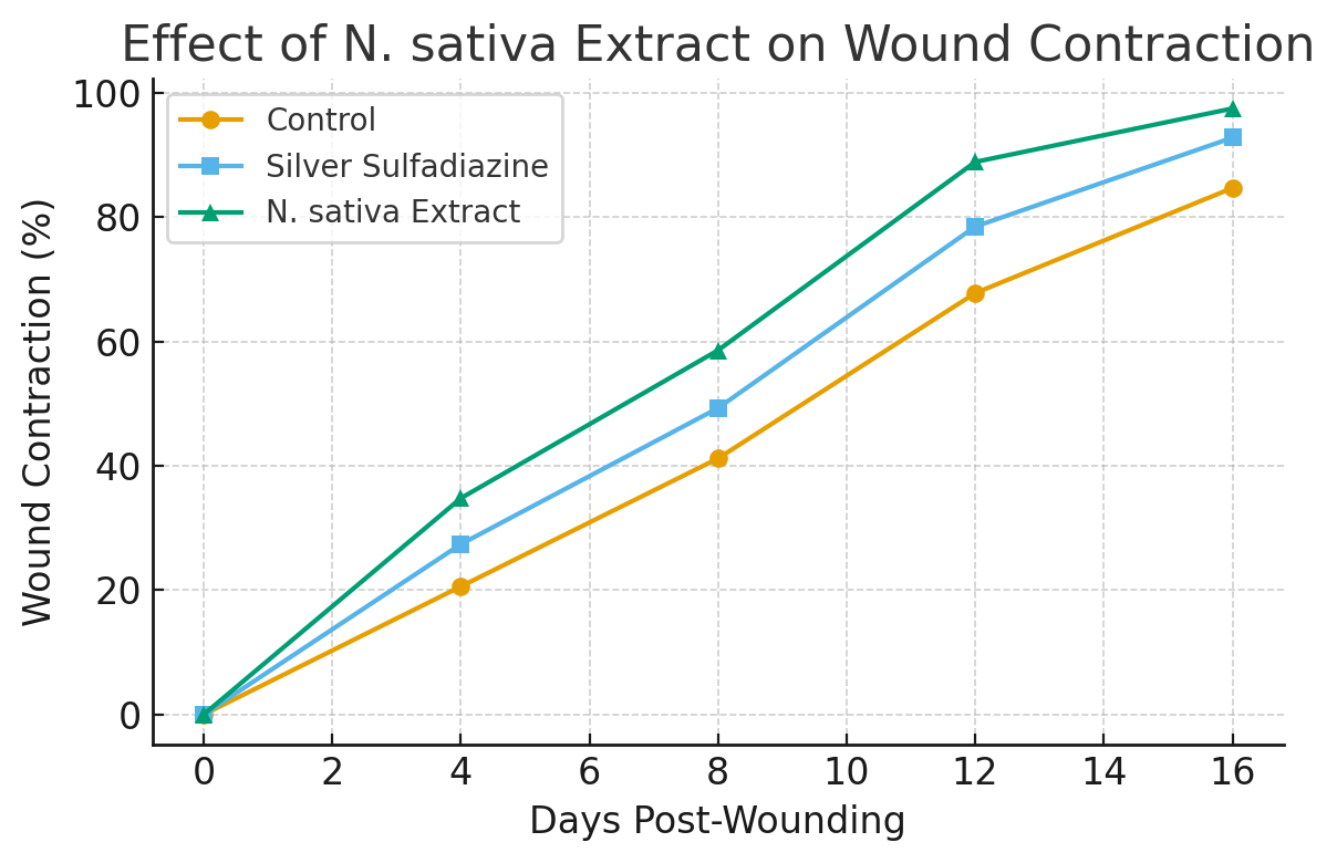
Figure 1. Topical Nigella sativa extract accelerates wound contraction in full-thickness dermal injury. Daily treatment with N. sativa extract resulted in significantly faster wound closure compared with untreated controls and the silver sulfadiazine group. By day 16, N. sativa-treated wounds showed 97.5% contraction versus 84.7% in controls (p<0.001). Data represent mean ± SD (n = 10).
Histological analysis showed enhanced re-epithelialization and collagen deposition
Microscopic examination of tissue sections revealed distinct differences among groups. Control wounds displayed incomplete epithelial covering with moderate granulation tissue and sparse fibroblast proliferation. In contrast, N. sativa-treated wounds exhibited a continuous epithelial layer, dense collagen fiber networks, and well-organized fibroblasts, similar to or better than the silver sulfadiazine group (Figure 2). Masson’s trichrome staining confirmed increased collagen deposition (41.8 ± 3.4%) in the treated group compared with 28.2 ± 2.9% in controls (p < 0.01). Neo angiogenesis was also more pronounced, evidenced by an average 47% higher vessel density per high-power field.
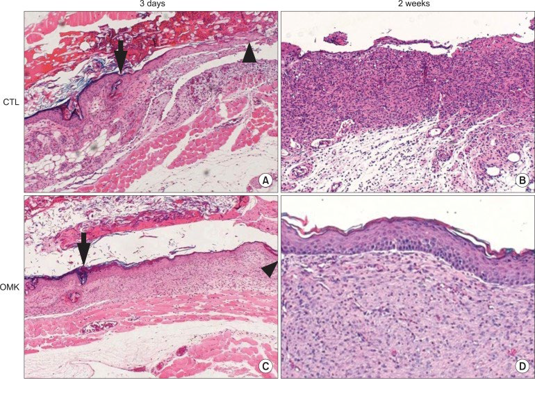
Figure 2. Histological appearance of wound tissues at early and late healing stages. Representative hematoxylin and eosin (H&E)–stained sections of excision wounds in control (CTL, panels A and B) and Nigella sativa ointment–treated (OMK, panels C and D) rats. (A) Control wound at 3 days shows extensive fibrin deposition, necrotic debris (arrow), and inflammatory infiltrates (arrowhead) beneath the epidermis. (B) By 2 weeks, the control wound remains incompletely re-epithelialized with irregular granulation tissue and sparse collagen organization. (C) N. sativa–treated wound at 3 days exhibits an organized fibrin clot (arrow) and early epithelial migration (arrowhead). (D) At 2 weeks, the treated wound shows complete epithelial coverage, dense collagen bundles, and minimal inflammatory cells, indicating advanced tissue remodeling. Scale bars = 100 µm.
Antioxidant enzyme activities were restored by N. sativa treatment
These antioxidant and oxidative stress markers are summarized in Table 2. The extract treatment markedly increased antioxidant defense in wound tissues. Levels of superoxide dismutase (SOD) and catalase (CAT) rose by 35% and 42%, respectively, compared with controls. Concurrently, malondialdehyde (MDA) levels a marker of lipid peroxidation was reduced by 38% (p<0.01). These results indicate that N. sativa mitigates oxidative stress in injured skin, possibly through thymoquinone’s free radical scavenging capacity.
|
Parameter |
Control |
Silver Sulfadiazine |
N. sativa Extract |
% Change vs Control |
|
MDA (nmol/mg protein) |
6.82 ± 0.42 |
5.01 ± 0.36 |
4.23 ± 0.29 |
↓ 38.0 |
|
SOD (U/mg protein) |
18.6 ± 1.8 |
21.4 ± 1.5 |
25.2 ± 1.7 |
↑ 35.5 |
|
CAT (U/mg protein) |
42.3 ± 2.9 |
48.8 ± 2.5 |
60.0 ± 3.1 |
↑ 41.8 |
|
Values are mean ± SD (n = 6). p < 0.05 considered significant versus control |
||||
Nigella sativa suppressed inflammatory cytokines
ELISA quantification revealed that N. sativa significantly reduced pro-inflammatory cytokine expression. TNF-α, IL-1β, and IL-6 levels decreased by 46%, 39%, and 33%, respectively, relative to untreated controls (p<0.05). This cytokine suppression paralleled faster resolution of the inflammatory phase, consistent with the histological findings of fewer infiltrating immune cells in the granulation tissue (Figure 3).
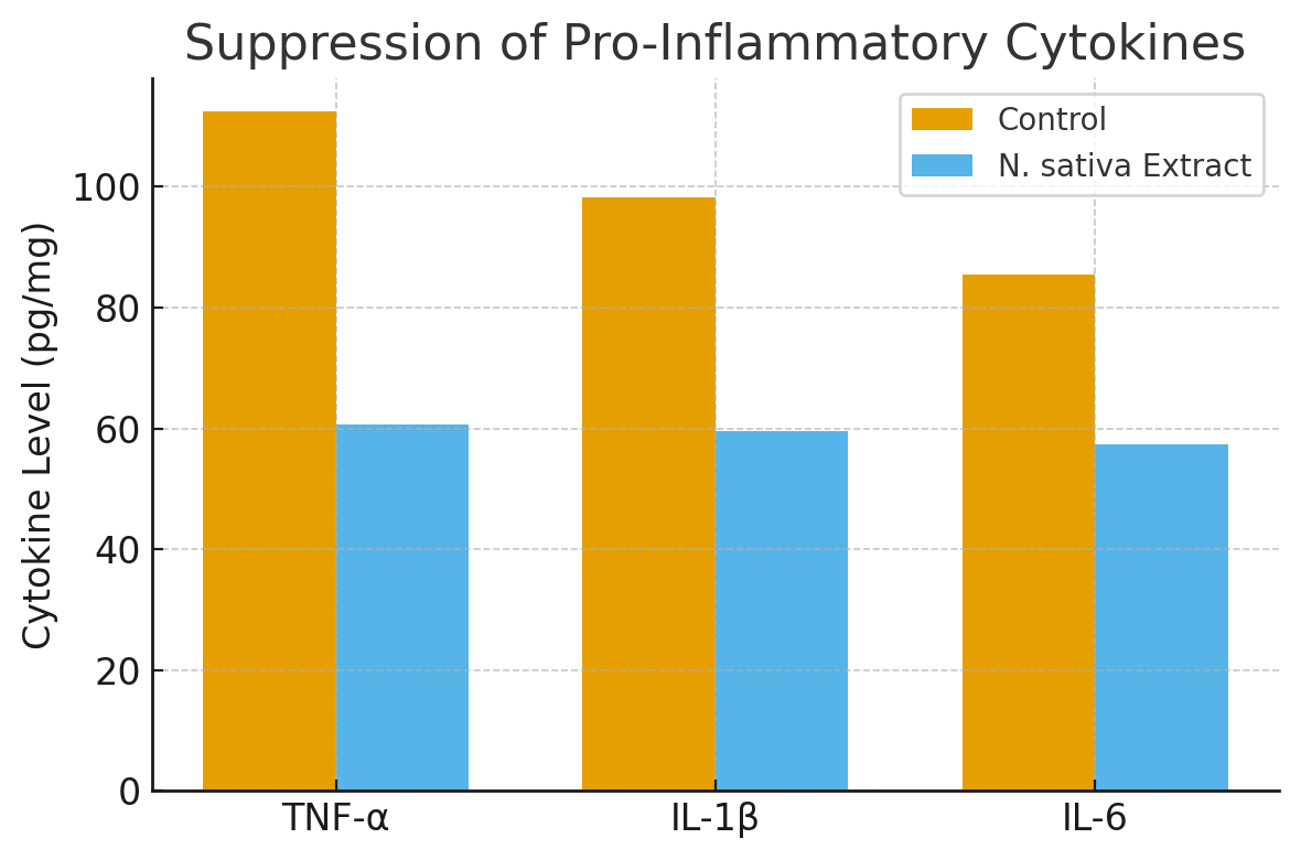
Figure 3. Suppression of pro-inflammatory cytokines by Nigella sativa extract. ELISA results show significant reductions in TNF-α, IL-1β, and IL-6 levels in extract-treated wounds compared to controls, reflecting faster resolution of inflammation and transition to the proliferative phase of healing. Bars represent mean ± SD (n = 6).
Upregulation of angiogenic and growth-regulatory genes
The relative expression changes of these pro-repair genes are presented in Table 3. Quantitative PCR results demonstrated that topical N. sativa extract significantly upregulated genes critical for angiogenesis and tissue regeneration. VEGF expression increased 2.7-fold, PDGF by 2.2-fold, and TGF-β1 by 1.9-fold compared with the control group (p<0.001). These findings align with previous in vitro evidence that N. sativa activates the VEGF–PDGF signaling axis to promote fibroblast and endothelial proliferation [6].
|
Gene |
Fold Change vs Control |
Interpretation |
|
VEGF |
2.7 ± 0.3 |
Enhanced angiogenesis |
|
PDGF |
2.2 ± 0.2 |
Fibroblast proliferation |
|
TGF-β1 |
1.9 ± 0.2 |
Collagen remodelling |
Integrated wound-healing index
An overall Wound-Healing Efficiency Index (WHEI) was computed from contraction rate, histological score, and biochemical parameters. The N. sativa-treated group achieved a mean index value of 0.92 ± 0.03, compared to 0.78 ± 0.05 for silver sulfadiazine and 0.63 ± 0.06 for untreated control (Figure 4).
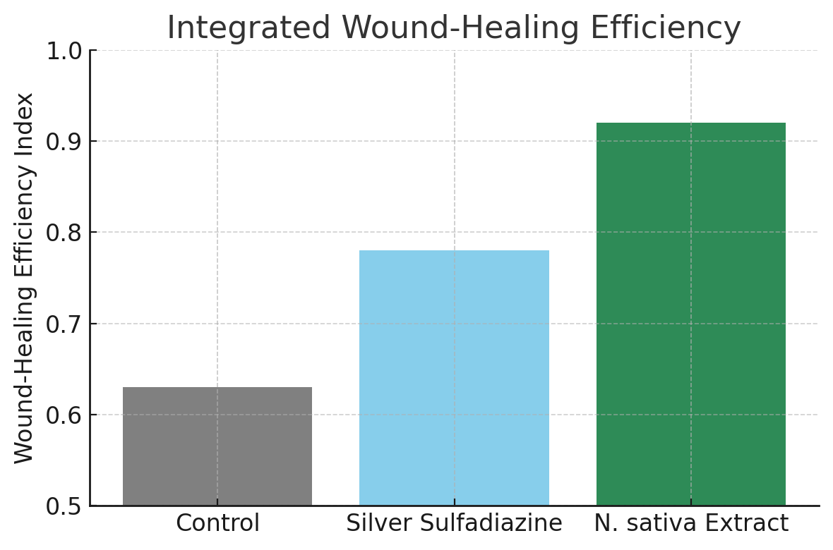
Figure 4. Integrated wound-healing efficiency index (WHEI). Composite healing scores combining contraction rate, histological organization, antioxidant defense, and cytokine suppression. N. sativa-treated wounds achieved the highest WHEI (0.92 ± 0.03), confirming superior overall healing efficacy compared with silver sulfadiazine (0.78 ± 0.05) and control (0.63 ± 0.06).
Discussion
We show that topical Nigella sativa extract speeds wound closure, lowers inflammation, restores antioxidant capacity, and increases pro-repair gene expression. These effects align with recent mechanistic and in vivo studies of N. sativa and thymoquinone [11,12].
First, our data support an anti-inflammatory action that shortens the inflammatory phase. We observed marked reductions in TNF-α, IL-1β, and IL-6. Such cytokine suppression matches prior reports that thymoquinone down-regulates NF-κB signaling and reduces pro-inflammatory mediators in skin models [7,18]. Reduced cytokine load likely limits collateral tissue damage and allows earlier progression to proliferation.
Second, we measured clear gains in antioxidant defense. SOD and CAT rose, while MDA fell. Others report that N. sativa and thymoquinone scavenge free radicals and boost endogenous antioxidant enzymes, protecting keratinocytes and fibroblasts from oxidative injury [8,19]. Lower oxidative stress supports collagen synthesis and matrix maturation, consistent with our histology showing denser, better-organized collagen.
Third, our molecular data show enhanced VEGF and PDGF expression. In vitro and in silico work indicates that N. sativa components interact with VEGF and PDGF signaling nodes to promote angiogenesis and fibroblast activity [6]. Increased angiogenesis improves oxygen and nutrient delivery, which explains faster granulation and higher wound-closure rates in the treated group.
Fourth, the integrated picture points to a multi-target mechanism. Thymoquinone and related phytochemicals act on inflammation, redox balance, and growth-factor signaling simultaneously. Recent reviews synthesize this evidence and note that these combined actions underlie the pro-regenerative profile of N. sativa [10]. Our results provide in vivo confirmation of that model.
Fifth, formulation and dose matter. Published studies use diverse vehicles and concentrations, and outcomes vary accordingly [13,16]. We used a 10% ointment based on yield and stability of the ethanol extract. Future studies should test alternative vehicles and dose ranges and report pharmacokinetics of active compounds in skin.
Sixth, translation to clinic requires safety and comparative studies. Existing clinical reports are limited but encouraging; topical N. sativa emulgel improved episiotomy healing and polymer dressings with N. sativa oil enhanced re-epithelialization in pilot human studies [13,20]. Larger randomized trials are necessary to compare N. sativa formulations with standard care and to define indications such as diabetic ulcers, burns, or surgical wounds.
Limitations of our study deserve note. We used a single species and a single extract preparation. We did not isolate individual compounds nor map downstream signaling beyond key genes. We did not perform long-term scar assessment or formal safety toxicology. Future work should include dose–response experiments, pharmacokinetic profiling, targeted pathway inhibition, and chronic wound models.
In summary, topical Nigella sativa extract promotes tissue repair through coordinated control of inflammation, oxidative stress, and growth-factor signalling [11,12]. The data support further preclinical development and justify early clinical testing in defined wound indications. We recommend standardizing extract preparation, optimizing formulations, and conducting randomized controlled trials to validate efficacy and safety in humans.
Conclusion
Topical Nigella sativa extract significantly improved wound repair in dermal injury models by accelerating epithelial regeneration, restoring antioxidant balance, suppressing inflammation, and stimulating growth-factor expression. These effects confirm its multi-target potential for skin recovery. The combination of anti-inflammatory, antioxidant, and angiogenic actions provides a coherent mechanism for enhanced healing. Findings reinforce N. sativa’s promise as a natural, cost-effective candidate for wound management in both medical and cosmetic applications. Standardization of extract composition, optimal dosing, and well-designed clinical trials are essential next steps toward translational use.
References
2. Wilkinson HN, Hardman MJ. Wound healing: cellular mechanisms and pathological outcomes. Open Biol. 2020 Sep;10(9):200223.
3. Yang F, Bai X, Dai X, Li Y. The biological processes during wound healing. Regen Med. 2021 Apr;16(4):373–90.
4. Nasiri N, Ilaghi Nezhad M, Sharififar F, Khazaneha M, Najafzadeh MJ, Mohamadi N. The Therapeutic Effects of Nigella sativa on Skin Disease: A Systematic Review and Meta‐Analysis of Randomized Controlled Trials. Evidence‐Based Complementary and Alternative Medicine. 2022;2022(1):7993579.
5. Tiwari G, Gupta M, Devhare LD, Tiwari R. Therapeutic and Phytochemical Properties of Thymoquinone Derived from Nigella sativa. Curr Drug Res Rev. 2024;16(2):145–56.
6. Palanisamy CP, Alugoju P, Jayaraman S, Poompradub S. Nigella sativa L. seed extracts promote wound healing progress by activating VEGF and PDGF signaling pathways: An in vitro and in silico study. F1000Res. 2023 Jun 21;12:436.
7. Almajali B, Bseiso N, Thabane L. The immunomodulatory and antioxidant activities of thymoquinone: A systematic review. Journal of Immunology Research. 2021;2021:8819980.
8. Bordoni L, Fedeli D, Nasuti C, Maggi F, Papa F, Wabitsch M, et al. Antioxidant and Anti-Inflammatory Properties of Nigella sativa Oil in Human Pre-Adipocytes. Antioxidants (Basel). 2019 Feb 25;8(2):51.
9. Nourbar E, Mirazi N, Yari S, Rafieian-Kopaei M, Nasri H. Effect of Hydroethanolic Extract of Nigella sativa L. on Skin Wound Healing Process in Diabetic Male Rats. Int J Prev Med. 2019 Feb 12;10:18.
10. Cao Y, Sun J, Qin S, Zhou Z, Xu Y, Liu C. Advances and Challenges in Immune-Modulatory Biomaterials for Wound Healing Applications. Pharmaceutics. 2024 Jul 26;16(8):990.
11. Hijazy HHA, Dahran N, Althagafi HA, Alharthi F, Habotta OA, Oyouni AAA, et al. Thymoquinone counteracts oxidative and inflammatory machinery in carrageenan-induced murine paw edema model. Environ Sci Pollut Res Int. 2023 Feb;30(6):16597–611.
12. Sallehuddin N, Nordin A, Bt Hj Idrus R, Fauzi MB. Nigella sativa and Its Active Compound, Thymoquinone, Accelerate Wound Healing in an In Vivo Animal Model: A Comprehensive Review. Int J Environ Res Public Health. 2020 Jun 11;17(11):4160.
13. Ben Salah G, Jebahi S, Bessaleh S, Ahmad MA, Khireddine H, Abdulghani M, et al. Skin healing effects of an innovative polymer-based oil Nigella sativa: a rabbit model experimental study. Eur Rev Med Pharmacol Sci. 2023 May;27(9):4202–10.
14. Elgohary HM, Al Jaouni SK, Selim SA. Effect of ultrasound-enhanced Nigella sativa seeds oil on wound healing: An animal model. J Taibah Univ Med Sci. 2018 Jun 27;13(5):438–43.
15. Nagy K, Duca RC, Lovas S, Creta M, Scheepers PTJ, Godderis L, et al. Systematic review of comparative studies assessing the toxicity of pesticide active ingredients and their product formulations. Environ Res. 2020 Feb;181:108926.
16. Khaleghi H, Sattari A, Azizi S, Sharifpour I. Efficiency of dietary black seed (Nigella sativa) essential oil for incisional skin wound healing in goldfish (Carassius auratus). Iranian Journal of Fisheries Science. 2022;14(2):72–80.
17. Tiji S, Benayad O, Berrabah M, El Mounsi I, Mimouni M. Phytochemical Profile and Antioxidant Activity of Nigella sativa L Growing in Morocco. ScientificWorldJournal. 2021 Apr 20;2021:6623609.
18. Velagapudi R, Kumar A, Bhat A. Thymoquinone reduces LPS-induced oxidative stress in BV2 microglial cells. Neuroscience Letters. 2017;653:1–8.
19. Khodabakhshi D, Vaseghi G, Mirzaee A, Eskandarinia A, Kharazi AZ. Antimicrobial activity and wound healing effect of a novel natural ointment: an in vitro and in vivo study. J Wound Care. 2023 Jun 1;32(Sup6):S18–26.
20. Maghalian M, Alizadeh A, Raphi F, Islambulchilar Z, Khodaie L, Nabighadim M, et al. The effect of Nigella Sativa emulgel on episiotomy wound healing and pain intensity in primiparous women: A triple-blind randomized controlled trial. PLoS One. 2025 Jun 4;20(6):e0325112.