

Abstract
Risk stratification for gastrointestinal stromal tumor (GIST) depends on postoperative mitotic index (MI), a histologic marker unavailable before surgery to guide neoadjuvant therapy and operative strategy. In 2020, Ren et al. constructed a routine CT nomogram to preoperatively identify localized primary GIST featured with high mitotic activity >5 mitoses per 50 high-power fields (>5/50 HPF), showing strong discriminative power in single-center training and internal validation cohorts. This commentary frames this imaging model within precision oncology paradigms, elaborating its clinical value while unpacking the key flaws of retrospective single-center prediction research. We further analyze the mismatches between macroscopic CT phenotypes and KIT/PDGFRA molecular profiles, specify strict clinical boundaries for the nomogram, and propose necessary translational validation workups. Although CT-based risk scores effectively streamline sarcoma MDT referrals, biopsy scheduling and intraoperative rupture prevention, they cannot substitute for the formal histopathology or mutational testing. Ultimately, visual CT nomograms only serve as auxiliary triage tools instead of main decision evidence in comprehensive GIST clinical management.
Keywords
Gastrointestinal stromal tumor, Mitotic index, Computed tomography, Nomogram, Preoperative risk stratification, Precision oncology, Sarcoma multidisciplinary team
Highlights
- Conventional CT morphological nomograms are auxiliary triage tools and cannot displace histopathology or KIT/PDGFRA sequencing for definitive GIST treatment decisions.
- Widespread molecular-morphology mismatches are present in GIST, and exclusive reliance on CT risk scores may lead to inappropriate imatinib and delayed avapritinib administration for PDGFRA exon 18 D842V-mutated tumors.
- Single-center retrospective design without external validation constrains the generalizability of the CT mitotic index prediction model proposed by Ren and colleagues.
- Strict clinical eligibility must be followed. The nomogram is only applicable to suspected primary localized GIST to facilitate MDT discussion, biopsy prioritization and surgical planning.
Introduction
Risk-adapted individualized treatment represents the core paradigm for localized gastrointestinal stromal tumor (GIST) clinical management, with all guideline-recommended therapeutic strategies determined by predicted malignant biological behavior of primary lesions [1]. Globally accepted risk stratification frameworks, including modified NIH criteria and AFIP pathological grading systems, depend on two core pathological indicators: maximum tumor diameter and mitotic index (mitotic figures per 50 high-power fields, HPF) [2]. Preoperative cross-sectional CT reliably quantifies tumor size, anatomical location, and macroscopic morphological features with excellent inter-examination reproducibility. However, definitive mitotic index quantification can only be completed via postoperative histopathological slide counting, creating a critical clinical bottleneck for upfront treatment planning [3].
This unmet clinical challenge creates a practical limitation in routine GIST care. The unconfirmed high mitotic risk prior to surgical resection constrains MDT risk assessment, targeted biopsy scheduling, and surgical technique design for preventing intraoperative tumor rupture. While endoscopic ultrasound (EUS)-guided or percutaneous core needle biopsy is generally safe for selected patients and preoperative tissue confirmation may alter treatment pathways, biopsy carries minor risks of tumor seeding and hemorrhage and small gastric GISTs may be directly managed via endoscopic resection without pre-operative biopsy [4,5]. The non-invasive preoperative imaging biomarkers for assessing mitotic risk would be of tangible clinical value for sarcoma patient triage.
Against this background, Ren and colleagues constructed a conventional CT feature-based nomogram to preoperatively identify patients with high mitotic index, providing a plausible low-cost, widely accessible preoperative risk prediction tool [6,7]. This commentary expands upon this single-center retrospective cohort research, assesses the translational advantage and inherent limitations of visual CT prediction models, compares conventional CT nomograms with emerging radiomics and liquid biopsy platforms, and establishes standardized clinical implementation boundary to guide safe, evidence-based real-world application of this predictive tool in precision GIST oncology.
The Contribution of the Focal Article
The landmark retrospective cohort study by Ren et al. enrolled 250 patients with pathologically confirmed primary localized GIST across a 5-year single-center dataset, randomly split at a 7:3 ratio into training cohort (n=176) and internal validation cohort (n=74) [8]. The univariate and multivariate binary logistic regression screening identified three independent CT imaging predictors of high mitotic index (>5/50 HPF): larger tumor size, intratumoral liquefaction and necrosis, and irregular tumor contour. The visual CT nomogram yielded an AUC of 0.851 in the training set and 0.836 in internal validation, demonstrating strong discriminative ability to distinguish high versus low mitotic risk GISTs using routine unenhanced and contrast-enhanced abdominal CT sequences available in all general radiology departments worldwide.
From surgical and medical oncology perspectives, the primary translational advantage of this model lies in its utilization of standard, qualitative CT morphological features without advanced post-processing, artificial intelligence reconstruction, or high-throughput radiomic feature extraction [9,10]. Each imaging biomarker carries clear biological underpinnings for aggressive mesenchymal tumor proliferation. Rapid tumor cell proliferation outpaces intralesional angiogenesis, leading to tissue hypoxia, liquefactive necrosis, and low-density non-enhancing areas on CT imaging. Irregular, lobulated tumor margins reflect asymmetric, invasive growth patterns, distinct from smooth, well-circumscribed borders observed in indolent, low-mitotic GIST subtypes [11]. In short, the three readily identifiable CT phenotypes enable radiologists to generate a preoperative numerical risk score without specialized software, streamlining rapid clinical risk triage before histology results.
Plausibly, the standard preoperative CT provides indirect, actionable surrogate markers of tumor proliferative kinetics, addressing the critical limitation of absent mitotic index data in pre-surgical patient evaluation [12,13]. However, robust discrimination statistics alone does not sufficiently justify standalone clinical decision-making, which requires layered clinical interpretation to avoid over-reliance on imaging surrogates.
Clinical Interpretation: Useful Signal but Not a Diagnostic Substitute
Despite favorable predictive performance metrics, clinicians have to interpret nomogram risk scores with strict bedside caution, with two clear clinical guardrails to prevent mismanagement.
First, preoperative CT morphological features could serve as quick triage aids rather than substitutes for histopathological mitotic quantification and molecular mutation testing. CT findings alone could hardly warrant neoadjuvant imatinib or other targeted therapies, as guideline-compliant neoadjuvant management requires histological validation plus KIT/PDGFRA genotyping to confirm GIST and rule out imatinib-resistant subtypes [14]. Similar to endoscopic risk stratification for early gastric cancer, CT-based scoring is not to replace postoperative pathology to assess lymphovascular invasion and metastatic potential [15].
Second, high nomogram scores prompt urgent sarcoma MDT consultation, refined biopsy schedules and adjusted surgical strategies in prevention of intraoperative tumor rupture, an event that by itself worsens prognosis and elevates disease stage [16,17]. This nomogram mainly functions to triage high-risk patients for specialized sarcoma management, instead of replacing tissue diagnosis.
Overreliance on CT nomograms is prone to notable clinical risks. For example, abdominal mesenchymal mimics (leiomyosarcoma, inflammatory myofibroblastic tumor) with necrotic CT features may falsely yield high mitotic risk scores insensitive to imatinib. For metastatic GIST, these CT models could hardly guide cytoreductive surgery or systemic therapy sequencing, as metastatic management strictly depends on tumor burden and molecular genotype instead of primary lesion imaging surrogates [18].
Methodological Considerations for Prediction-Model Studies
To facilitate clinical adoption of this CT nomogram, we assessed its design per TRIPOD+AI guidelines to highlight critical methodological limitations [19].
Single-center retrospective cohort and internal validation bias
The retrospective single-center design inherently limits generalizability. Internal split-sample validation using uniform local CT protocols tends to overestimate real-world AUC, and multi-center external validation across varied equipment and populations is required to verify stable predictive performance.
Statistical modeling risks of overfitting
Stepwise logistic regression relying on p-values and AIC renders high overfitting risks for moderate cohorts. Standardized reporting of missing data and incomplete follow-up is also absent, a critical transparency shortfall [20].
Incomplete calibration and decision curve evaluation
High AUC does not guarantee clinical value, and practical models rely on validated calibration, inter-rater consistency and net clinical benefit [21]. The Hosmer-Lemeshow test is sample-size dependent and fails to assess full-range risk calibration. Subsequent validations should report complete calibration metrics and DCA to quantify clinical utility [22].
DCA thresholds demand clinical trade-offs: high-risk cutoffs balance detection of aggressive GISTs against unnecessary biopsy and overtreatment [23]. Qualitative CT features like irregular tumor margins are subject to radiologist judgment. Though consistent within this single center, inter-rater Kappa across general radiologists was unreported, restricting external applicability [24,25].
Molecular and Translational Context
When implementing CT nomograms for precision GIST management clinicians should account for tumor genomic drivers. Approximately 85% of sporadic GISTs harbor active KIT/PDGFRA mutations that determine proliferative potential and therapeutic response [26,27]. Mutation subtypes bear a non-linear relationship with mitotic index, and conventional CT morphological features fail to visualize subpixel molecular alterations underlying tumor progression.
Risk stratification relying on CT findings without genotyping frequently leads to misjudgment. Large necrotic gastric GISTs typically generate elevated nomogram risk scores, yet PDGFRA exon 18 D842V-mutated tumors exhibit primary imatinib resistance [28]. Administering neoadjuvant imatinib based solely on CT nomogram results delays avapritinib therapy and compromises patient survival outcomes [29,30]. Imaging biomarkers and molecular pathology serve as complementary, non-interchangeable diagnostic modalities.
While advanced radiomics and radiogenomics can extract subtle imaging phenotypes correlated with KIT/PDGFRA variants, these quantitative imaging techniques remain investigational and lack formal guideline endorsement [31]. Conventional CT nomograms cannot substitute mandatory molecular testing prior to systemic therapy in standard clinical workflows.
Clinical Implementation and Boundaries
This nomogram has strict clinical application limits. It benefits only suspected localized primary GISTs to guide MDT consultation, biopsy and surgery planning, with its utility determined by safe clinical decision improvement [32].
Ren’s cohort mainly consisted of gastric GISTs (76.7% of training set) [8]. Biopsy and neoadjuvant therapy for gastric lesions rely largely on tumor size and location, and low-risk cases merely need endoscopic resection and surveillance [33–35]. By contrast, small rectal or duodenal GISTs often demand high-morbidity radical operations like abdominoperineal or pancreaticoduodenal resection [36,37]. For extragastric GISTs, surgical planning prioritizes anatomy and operative risks instead of CT-predicted mitotic index, so the nomogram only serves as supplementary evidence in individualized MDT decisions [38].
Future directions
Four validation and comparative studies are necessary before routine clinical use of this CT nomogram.
- It calls for multi-center external validation covering various CT devices, contrast protocols and populations, with standardized reporting of calibration metrics and DCA curves [39]. Similar disparities in imaging access and diagnostic consistency across demographic subgroups have been widely documented in breast cancer care, underscoring the need for population-diversified model validation [40].
- It needs to integrate CT descriptive standards for necrosis and irregular margins to cut inter-reader variation and boost Kappa reliability in community radiology [41]. As unstandardized intraoperative endocrine assessment brings large observer bias, uniform abdominal tumor CT terminology is essential [42].
- It requires head-to-head comparisons with radiomic machine learning and cfDNA liquid biopsy assays [43]. Currently, cfDNA molecular testing for localized GIST remains investigational and cannot replace tissue-based genotyping standards [44].
- It demands for prospective trials to evaluate real-world outcomes, including intraoperative rupture, MDT efficiency, overall survival and cost-effectiveness of nomogram-based risk stratification [45].
For illustrating the auxiliary role of CT nomograms in standardized GIST preoperative management and for educational purpose, we summarize the complete clinical decision-making workflow as shown in Figure 1.
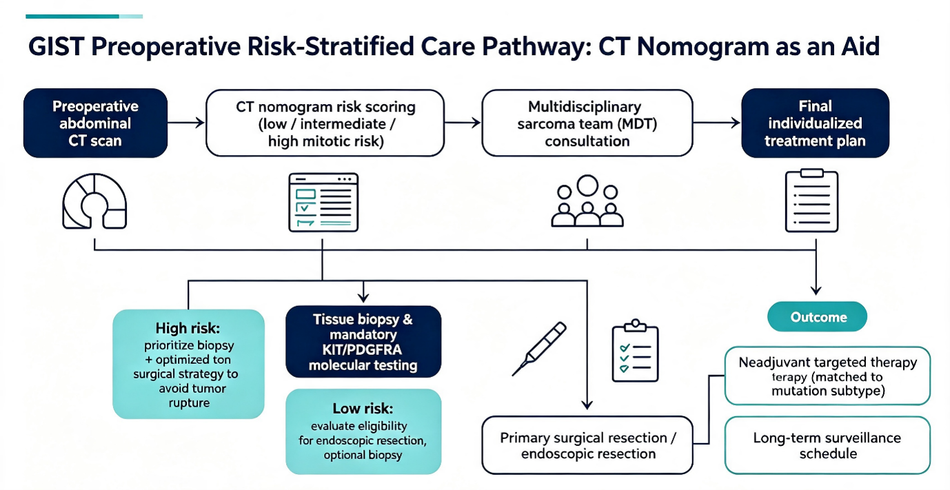
Figure 1.
Conclusions
Ren’s CT nomogram is an affordable preoperative triage tool for localized primary GIST, using three routine CT features (large size, intratumoral necrosis, irregular margins) to predict high mitotic risk. It shows plausible discrimination in single-center internal validation and offers biological surrogates to optimize surgery and sarcoma MDT scheduling.
However, this morphology-based nomogram cannot serve as an independent clinical decision tool due to methodological, molecular and anatomical constraints. It provides the auxiliary evidence and would further be combined with postoperative histopathology, mandatory KIT/PDGFRA genotyping and full sarcoma MDT assessment. Further multi-center external validation and prospective trials are necessary to define its clinical value in integrated GIST precision oncology.
Disclosures
Conflict of interest statement
All authors declare no conflicts of interest for this invited commentary manuscript.
Funding statement
This commentary received no dedicated external funding support apart from the Wu Jieping Medical Foundation grant supporting the original focal CT nomogram study. Wu Jieping Medical Foundation, Grant No. 320.6750.2022-3-41.
Author contributions
Yonghong Dong: Conceptualization, manuscript critical revision, corresponding author supervision; Junjie An: Literature retrieval, full manuscript drafting; Yingzheng Ren: Original focal study data interpretation, statistical model analysis; Yang Yang: Clinical oncology workflow validation, molecular pathology discussion revision. All authors approved the final submitted manuscript.
Data availability statement
No novel patient clinical or imaging datasets were generated or analyzed during the preparation of this commentary. All clinical model parameters, cohort characteristics, and statistical performance metrics discussed are sourced from the cited original published focal study.
References
2. Serrano C, Martín-Broto J, Asencio-Pascual JM, López-Guerrero JA, Rubió-Casadevall J, Bagué S, et al. 2023 GEIS Guidelines for gastrointestinal stromal tumors. Ther Adv Med Oncol. 2023 Aug;15:17588359231192388.
3. Barat M, Pellat A, Dohan A, Hoeffel C, Coriat R, Soyer P. CT and MRI of Gastrointestinal Stromal Tumors: New Trends and Perspectives. Can Assoc Radiol J. 2024 Feb;75(1):107–17.
4. Li J, Xu D, Huang WF, Hong SK, Zhang JY. Efficacy and Safety of Endoscopic Resection for Gastric Gastrointestinal Stromal Tumors Originating from the Muscularis Propria. Dig Dis Sci. 2024 Jun;69(6):2184–92.
5. Seven G, Arici DS, Senturk H. Correlation of Endoscopic Ultrasonography Features with the Mitotic Index in 2- to 5-cm Gastric Gastrointestinal Stromal Tumors. Dig Dis. 2022;40(1):14–22.
6. Ren C, Wang S, Zhang S. Development and validation of a nomogram based on CT images and 3D texture analysis for preoperative prediction of the malignant potential in gastrointestinal stromal tumors. Cancer Imaging. 2020 Jan 13;20(1):5–14.
7. Lin JX, Wang FH, Wang ZK, Wang JB, Zheng CH, Li P, et al. Prediction of the mitotic index and preoperative risk stratification of gastrointestinal stromal tumors with CT radiomic features. Radiol Med. 2023 Jun;128(6):644–54.
8. Yingzheng R, Linlin J, Yang Y, Junjie A, Yonghong D. Study on the predictive value of preoperative CT features for the mitotic index of GIST based on the nomogram. Sci Rep. 2025 Mar 13;15(1):8627.
9. Mazzei MA, Cioffi Squitieri N, Vindigni C, Guerrini S, Gentili F, Sadotti G, et al. Gastrointestinal stromal tumors (GIST): a proposal of a "CT-based predictive model of Miettinen index" in predicting the risk of malignancy. Abdom Radiol (NY). 2020 Oct;45(10):2989–96.
10. Xu J, Zhou J, Wang X, Fan S, Huang X, Xie X, et al. A multi-class scoring system based on CT features for preoperative prediction in gastric gastrointestinal stromal tumors. Am J Cancer Res. 2020 Nov 1;10(11):3867–81.
11. Wei SC, Xu L, Li WH, Li Y, Guo SF, Sun XR, et al. Risk stratification in GIST: shape quantification with CT is a predictive factor. Eur Radiol. 2020 Apr;30(4):1856–65.
12. Zhang X, Bai L, Wang D, Huang X, Wei J, Zhang W, et al. Gastrointestinal stromal tumor risk classification: spectral CT quantitative parameters. Abdom Radiol (NY). 2019 Jul;44(7):2329–36.
13. Grazzini G, Guerri S, Cozzi D, Danti G, Gasperoni S, Pradella S, et al. Gastrointestinal stromal tumors: relationship between preoperative CT features and pathologic risk stratification. Tumori. 2021 Dec;107(6):556–63.
14. van der Burg SJC, van de Wal D, Roets E, Steeghs N, van Sandick JW, Kerst M, et al. Neoadjuvant Imatinib in Locally Advanced Gastrointestinal Stromal Tumors (GISTs) is Effective and Safe: Results from a Prospective Single-Center Study with 108 Patients. Ann Surg Oncol. 2023 Dec;30(13):8660–68.
15. Souza WP, Pereira MA, Cardili L, Zilberstein B, Ribeiro-Junior U, Ramos MFKP. Evaluation of the endoscopic cure criteria in patients undergoing surgery for early gastric cancer. J Surg Oncol. 2024 Sep;130(4):743–9.
16. Judson I, Jones RL, Wong NACS, Dileo P, Bulusu R, Smith M, et al. Gastrointestinal stromal tumour (GIST): British Sarcoma Group clinical practice guidelines. Br J Cancer. 2025 Jan;132(1):1–10.
17. Nishida T, Gotouda N, Takahashi T, Cao H. Clinical importance of tumor rupture in gastrointestinal stromal tumor. J Dig Dis. 2024 Sep-Oct;25(9-10):542–9.
18. Liu DN, Jia WW, Wang HY, Wu JH, Li CP, Hao CY. Cytoreductive surgery offers prognostic benefits in metastatic gastrointestinal stromal tumors with generalized progression following imatinib therapy: a single institute retrospective study. BMC Surg. 2023 Jul 4;23(1):189–97.
19. Collins GS, Moons KGM, Dhiman P, Riley RD, Beam AL, Van Calster B, et al. TRIPOD+AI statement: updated guidance for reporting clinical prediction models that use regression or machine learning methods. BMJ. 2024 Apr 16;385:e078378.
20. Ibi Y, Omori T. Cure rate estimation with insufficient follow-up: A median-based bootstrap correction approach. PLoS One. 2026 Mar 12;21(3):e0344669.
21. O’Reilly E, Kurakovas T. The Independence of Discrimination and Calibration in Clinical Risk Prediction: Lessons from a Multi-Timeframe Diabetes Prediction Framework. medRxiv. 2026 Feb 14:2026–02. doi:10.64898/2026.02.12.26346147.
22. Lee WJ, Asghar MS, Park R, Won SH, Shahzad M, Shimshak T. Externally Validated Machine Learning Models for 30-/90-/180-Day Unplanned Readmission After Head and Neck Cancer Hospitalizations in the United States. JCO Clin Cancer Inform. 2026 Apr;10(2):e2500335.
23. Vickers AJ, Elkin EB. Decision curve analysis: a novel method for evaluating prediction models. Med Decis Making. 2006 Nov-Dec;26(6):565–74.
24. Doyle MC, Timpone VM. Patient-Friendly Summary of the ACR Appropriateness Criteria®: Myelopathy. J Am Coll Radiol. 2026 Mar;23(3):483.
25. Zhang Y, Zhang H, Li D, Yuan L, Deng L, Han T, et al. Contrast-enhanced CT-derived extracellular volume fraction in preoperative prediction of tumor-stroma ratio in colorectal cancer. Abdom Radiol (NY). 2026 Feb;51(2):639–49.
26. Miettinen M, Lasota J. Gastrointestinal stromal tumors: review on morphology, molecular pathology, prognosis, and differential diagnosis. Arch Pathol Lab Med. 2006 Oct;130(10):1466–78.
27. Boye K, Gorunova L, Gunawan B, Hompland I, Sander B, Panagopoulos I, et al. Genomic Complexity as a Biomarker to De-Escalate Adjuvant Imatinib Treatment in High-Risk Gastrointestinal Stromal Tumor. JCO Precis Oncol. 2023 Feb;7:e2200351.
28. Casali PG, Blay JY, Abecassis N, Bajpai J, Bauer S, Biagini R, et al. Electronic address: clinicalguidelines@esmo.org. Gastrointestinal stromal tumours: ESMO-EURACAN-GENTURIS Clinical Practice Guidelines for diagnosis, treatment and follow-up. Ann Oncol. 2022 Jan;33(1):20–33.
29. Xie Z, Zhang Q, Zhang R, Zhao Y, Zhang W, Song Y, et al. Identification of D842V mutation in gastrointestinal stromal tumors based on CT radiomics: a multi-center study. Cancer Imaging. 2024 Dec 20;24(1):169–77.
30. Beecroft JR, Brar S, Feng X, Hamilton T, Han-Lee C, Henning JW, et al. Pan-Canadian consensus recommendations for GIST management in high- and low-throughput centres across Canada. Ther Adv Med Oncol. 2024 Aug 2;16:17588359241266179.
31. Galluzzo A, Boccioli S, Danti G, De Muzio F, Gabelloni M, Fusco R, et al. Radiomics in gastrointestinal stromal tumours: an up-to-date review. Jpn J Radiol. 2023 Oct;41(10):1051–61.
32. Tang P, Li M, Kang Y, Wu X. Development of an Individualized CT-based Radiomics Nomogram for Preoperative Differentiation Between Gastric Stromal Tumors and Gastric Leiomyomas. J Vis Exp. 2026 Feb 3;(228).
33. Gao J, Liu Z, Liu X, Shu X, Zhu Y, Chen Y, Zeng C. Follow-up analysis and research of very low-risk and low-risk gastrointestinal stromal tumors after endoscopic resection. Sci Rep. 2024 Aug 2;14(1):17872.
34. Chen T, Xu L, Dong X, Li Y, Yu J, Xiong W, et aal. The roles of CT and EUS in the preoperative evaluation of gastric gastrointestinal stromal tumors larger than 2 cm. Eur Radiol. 2019 May;29(5):2481–9.
35. Jia X, Wan L, Chen X, Ji W, Huang S, Qi Y, et al. Risk stratification for 1- to 2-cm gastric gastrointestinal stromal tumors: visual assessment of CT and EUS high-risk features versus CT radiomics analysis. Eur Radiol. 2023 Apr;33(4):2768–78.
36. Shen Z, Chen P, Du N, Khadaroo PA, Mao D, Gu L. Pancreaticoduodenectomy versus limited resection for duodenal gastrointestinal stromal tumors: a systematic review and meta-analysis. BMC Surg. 2019 Aug 28;19(1):121.
37. Mangana O, Liccardo F, Sepe E, Celentano V. Revisional ileoanal pouch surgery with in situ pouch augmentation for recurrent pouch inlet stricture. Colorectal Dis. 2025 Jan;27(1):e17241.
38. Hølmebakk T, Wiedswang AM, Meza-Zepeda LA, Hompland I, Lobmaier IVK, Berner JM, et al. Integrating Anatomical, Molecular and Clinical Risk Factors in Gastrointestinal Stromal Tumor of the Stomach. Ann Surg Oncol. 2021 Oct;28(11):6837–45.
39. Jiang Q, Fu Y, Zhang X, Zhang P, Gao Z, Zhang J, et al. Distinguishing very high-risk patients among high-risk gastrointestinal stromal tumor cases: development and validation of a nomogram based on a multicenter population-based retrospective cohort study. Ann Med. 2025 Dec;57(1):2520896.
40. Arrey EN, GoPaul D, Anderson D, Okoli J, McKenzie-Johnson T. Addressing Breast Cancer Disparities: A Comprehensive Approach to Health Equity. J Surg Oncol. 2024 Dec;130(7):1483–89.
41. Lee T, Kim H, Lee S, Chae S, Kahn CE , Jr, You SC, et al. Large Language Model-Generated Expansion of the RadLex Ontology: Application to Multinational Datasets of Chest CT Reports. AJR Am J Roentgenol. 2026 May;226(5):e2534243.
42. Vetter D, Triponez F. Near-infrared fluorescence techniques to reduce hypocalcaemia after thyroidectomy. Br J Surg. 2023 Nov 9;110(12):1590–3.
43. Zheng T, Du J, Yang L, Dong Y, Wang Z, Liu D, et al. Evaluation of risk classifications for gastrointestinal stromal tumor using multi-parameter Magnetic Resonance analysis. Abdom Radiol (NY). 2021 Apr;46(4):1506–18.
44. Sun J, Li J, He Y, Kang W, Ye X. Identification and validation of protein biomarkers for predicting gastrointestinal stromal tumor recurrence. Comput Struct Biotechnol J. 2024 Feb 22;23:1065–75.
45. Xu SJ, Zhang SY, Dong LY, Lin GS, Zhou YJ. Dynamic survival analysis of gastrointestinal stromal tumors (GISTs): a 10-year follow-up based on conditional survival. BMC Cancer. 2021 Nov 1;21(1):1170.