

Abstract
Both genetic and environmental factors contribute to the development of autoimmune disease and allergy. Increased disease prevalence is associated with living in higher latitudes, and decreased exposure to sunlight as a child. Whilst ultraviolet radiation has known anti-inflammatory properties, its ability to influence the development of immune tolerance during childhood years is not established. Regulatory T-cells (Tregs) play a major role in suppressing excessive immune reactions in both autoimmunity and allergy, and a reduction in Treg capacity can predispose an individual to disease. Levels of Tregs are highest at birth and remain high until puberty, with this being the time when the developing immune system is exposed to antigen and T-cells ‘learn’ what to react to. Circulation of Tregs and interaction with dendritic cells is controlled by the circadian light pattern via cortisol and intracellular circadian genes. Photoperiod and visible light intensity, which are reduced in winter at high latitudes, influence cortisol levels and circulation and production of Tregs. This review provides a model of the circadian mechanism whereby reduced light gives rise to reduced levels of Tregs and reduced interaction with antigen, which, during the critical developmental period, leads to a reduced pool of ‘trained’ Tregs. Examples from human and diurnal animal studies are provided.
Keywords
Autoimmune, Environment, Latitude, T-regulatory, Photoperiod
The Latitude Gradient of Allergy and Autoimmune Disease
Autoimmune diseases such as Multiple Sclerosis (MS), Rheumatoid Arthritis (RhA) and Type 1 Diabetes (Diab T1) arise when the immune system mistakenly mounts an attack on self-tissue, resulting in damage and dysfunction. Allergy is an over-reaction of the immune system to an environmental allergen such as food or pollen, potentially resulting in a systemic inflammatory reaction affecting the airways or skin. Both autoimmune diseases and allergies have a latitudinal distribution, with higher prevalence in higher latitudes, that is towards the poles [1,2]. In the southern hemisphere, in Australia (latitude spread of 10°- 43°) and New Zealand (latitude 29°-52°), the prevalence for MS for people who grew up in the southern zones of the countries is 2–3 times that for those who grew up in the north [3,4], Figure 1. Similarly, the risk for eczema is at least twice that for children in the south of Australia than for those in the north [5], whilst the rate of peanut allergy in 8–9-year-olds is six times higher in the south compared to the north [5], Figure 1. Rates of anaphylaxis were significantly higher in the south of Australia and Chile than the north [6,7]. In the United States, a latitudinal variation in the incidence of inflammatory bowel disease in women was observed with higher rates in the north [8]. A study of nurses in the US demonstrated an association of higher prevalence of Rheumatoid Arthritis with decreased Ultraviolet (UV) exposure, occurring generally more in northern/higher latitudes where there is lower UV [9]. A study of boys in Australia who developed Type 1 Diabetes by the age of 15 demonstrated that those who had lower UV exposure, generally in the more southern/higher latitudes, had a greater risk of disease [10]. These are just a few of many examples of the latitude gradient of disease, raising the question of how higher latitude predisposes to disease.
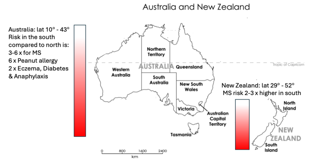
Figure 1. Latitude gradient of autoimmune disease and allergy in Australia and New Zealand, risk of autoimmune disease and allergy is greater in higher latitudes in the south.
Association of Disease with Reduced Sunlight Exposure: Lack of Daylight, Not Vitamin D
It is generally agreed that the differences in prevalence and risk of disease at high and low latitudes is associated with the amount of sunlight that the person has been exposed to up until their early teens [4], with lower exposure associated with greater risk of disease [2]. However, there is an ongoing debate over the ‘type’ or frequency of wavelength of sunlight that is responsible for the phenomenon, and the mechanisms involved. The spectrum of sunlight that influences biological functions range from Ultraviolet (UV) A (320-400 nm) & B (280-320 nm) through visible light (400-700 nm) to infrared radiation (>700 nm), Figure 2. Ultraviolet B radiation has been touted as the likely frequency of sunlight that is responsible for immune changes, originally due to its ability to produce previtamin D3, the precursor to vitamin D in the skin. Studies of associations of vitamin D levels and risk of allergy or autoimmune disease are common [11], but it is more likely that vitamin D levels are acting as a proxy for daylight exposure. Clinical trials of Vitamin D have not been consistently successful in treatment of autoimmune disease, and there are no clear mechanisms of how vitamin D deficiency in childhood leads to disease. That is, it has not been demonstrated that Vitamin D can directly alter populations and function of relevant immune cells to the extent that would confer predisposition to disease. It is now apparent that UV radiation can stimulate the production of an array of anti-inflammatory factors [12,13]. Direct UV has successfully been used to treat skin conditions due to its anti-inflammatory properties. T-helper 17 cells (Th17), an effector cell implicated in inflammatory and autoimmune conditions, are influenced by UV, as demonstrated in dermatological studies [14]. A study of healthy women in the south of Australia demonstrated an inverse correlation between number of hours spent outside and Th17 levels, suggesting Th17 decreases with short bursts of sunlight [15]. There is little known of how a lack of UV during childhood influences or primes the number or the function of effector immune cells which later contributes to disease onset. As the effect of UV on the immune system and inflammatory mediators has been discussed in other reviews [16] the focus of this paper will focus on the effect of visible light on immune development.
In regard to the link between decreased sunlight exposure in childhood and disease, the questions we were interested in were what type of sunlight are children not getting sufficient exposure to, and what aspect of the immune system is being primed or not being sufficiently developed over multiple years, to lay the foundation for lack of tolerance to self or allergens. To understand which frequency of solar radiation influences the development of the immune system prior to the onset of a condition, it is important to understand normal immune development including acquisition of immune tolerance. A deficiency in a normal developmental pathway would potentially provide a mechanism for disease predisposition. The scope of this review is to focus on the link between the visible light spectrum, circadian factors and regulatory T-cells, a major player in allergy and autoimmunity. The following sections provide an overview of regulatory T-cells, their circadian control and relevance to immune dysfunction.
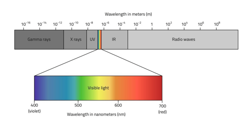
Figure 2. Light spectrum, with visible light wavelength ranging from 400-700nm.
Role of Regulatory T-cells in Disease
Regulatory T-cells, Tregs, recognize self-tissue and allergen and act to control or suppress excessive immune reactions. Tregs can employ various mechanisms against various immune players such as cell-to-cell interaction, release or inhibition of soluble mediators, or metabolic disturbance to reduce an immune reaction [17]. Tregs allow humans and other species to be ‘tolerant’ of our own tissue or innocuous stimulants. When Tregs are inadequate or their function goes awry, autoimmunity or allergy can arise. Indeed, lower levels of Tregs have been observed in young MS patients [18,19], other autoimmune diseases [20], in allergy [21–25] and are associated with disease severity in young RhA patients [26]. Lower Treg levels prior to, or in the early course of disease are of particular interest, as Treg levels may change with disease progression. Tregs have been demonstrated as being not only heterogeneous but highly plastic [17], and able to lose their regulatory ability in an inflammatory milieu. Altered Treg function has been observed in numerous autoimmune conditions, with the specific mechanism varying between conditions [27]. An animal model of dysfunctional FoxP3, the hallmark transcription factor of Tregs, results in ‘scurfy’, a severe autoimmune condition [28], and the human equivalent, immunodysregulation polyendocrinopathy enteropathy X-linked syndrome (IPEX syndrome) results in allergy and autoimmunity [17], illustrating the importance of Tregs to prevent autoimmune disease. A factor that reduces the normal production or circulation of Tregs would potentially predispose an individual to having a lower tolerance threshold and could possibly contribute to disease.
Early Life Development of Regulatory T-cells (Tregs): Influence of Light
Naïve Tregs are produced in the thymus as early as during fetal development [29] and the levels in newborns remain at their highest levels of life for the next few months, [30–34], Figure 3. By the first-year levels decrease slightly and then plateau until puberty [35,36]. After puberty there is a moderate decline in levels [35]. Interestingly, the output of new cells from the thymus is associated with the month of birth according to season [37], indicating that light exposure to the mother influences the production of new T-cells of the fetus. In relation to this there are reports of the month of birth being linked to higher risk of developing certain autoimmune diseases [38]. The critical period for naïve T-regs to recognize their cognate antigen and transition to memory Tregs is during infancy and childhood, when they are at their highest levels. If Tregs are not exposed to self or foreign antigen during this period, the potential to stop an unwanted reaction later in life will be reduced.
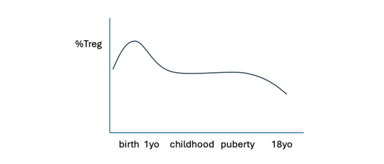
Figure 3. Change in %Treg during childhood and adolescence. Treg levels are highest around birth, and the first year, plateauing until puberty, after which the levels decrease.
Evidence of Seasonal Variation in Circulation of Tregs from Human Studies
The circulation of Tregs through the body occurs in a circadian rhythm [39]. Tregs begin to egress from the lymph nodes, the thymus and bone marrow out into the peripheral blood system from late morning, with levels increasing during the day and evening with highest levels being in the early hours of the morning around 2 AM, Figure 4. The timing and degree of circulation of Tregs varies according to the light-dark photoperiod which changes according to the season, with the greatest change being at the highest latitudes [2]. It was demonstrated that the evening circulation of T-regulatory cells was lower in winter compared to summer in pubescent girls and their mothers who lived at a moderately high latitude at the southern tip of Australia, where the daylight length is six hours less in winter than in summer [15]. In England, it was demonstrated that the morning levels of T-regulatory cells are lowest mid-winter and highest in summer in adult males and were associated with cortisol levels [40]. Studies in Scotland and Austria also demonstrated higher Tregs during summer months compared to winter, and this was independent of Vitamin D [41,42]. The results from these studies combined indicate that in the short daylengths of winter, there are lower levels of Tregs circulating in the body during the day and early evening compared to summer, where the longer daylengths result in higher levels of circulating Tregs, Figure 4. This decrease in circulation of Treg cells in winter in children during the critical developmental period has implications for regulating the immune system either against allergens as a child, or against self-tissue later in life.
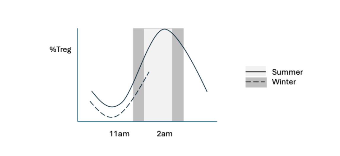
Figure 4. Circadian and seasonal pattern of Tregs. Tregs circulate in a circadian rhythm, with levels lower in the short daylight photoperiod in winter compared to summer.
Circadian Rhythm Controlled by Cortisol via HPA and Cellular Clocks
Treg migration out of lymph nodes, the thymus and bone marrow into the blood system is dependent on the circulating levels of cortisol, a circadian hormone [43–46]. In addition to having a circadian rhythm, cortisol has been demonstrated to have a circannual production profile, with concentrations being higher in winter than summer, with levels increasing with latitude, Figure 5. In Australia’s mainland, it was demonstrated that residents living above latitude 25° have higher cortisol levels in winter than summer, and those living in the most southern state (highest latitude) have the highest levels of cortisol in winter and summer [47]. Studies of seasonal cortisol levels at similar or higher latitudes in Israel, England and Sweden also demonstrate that the disparity in levels between summer and winter increases with increasing latitude [48]. This disparity provides a potential explanation of why there is a seasonal difference in circulating Treg levels. The following describes how light and cortisol control circadian clock genes which regulate circulation of T-cells.
Cortisol production is regulated by light via the Hypothalamic Pituitary Adrenal (HPA) Axis, Figure 6. Light entering the eye activates the Supra chiasmic nucleus (SCN), which in turn activates the HPA axis which results in the production of cortisol from the adrenal glands. Cortisol exerts its effect on immune cells, such as Tregs, via corticoid receptors which activate internal cellular clock genes such as ‘Clock’ (Circadian locomotor output cycles kaput) and Bmal1 (Brain muscle ARNT like 1). The genes operate in a negative feedback loop, whereby increased levels of circulating hormones will cause downregulation of the genes via expression of the heterodimers Per ½ (period) and Cry ½ (Cryptochrome). These circadian genes have been demonstrated to influence the circadian timing of transcriptional programs related to migration receptor expression [49,50] which will be explained in more detail below. Interestingly, circadian regulation has also been shown to be involved in T-cell differentiation of CD4+ cells toward Th1, Th2, Th17, the effector T-cells implicated in autoimmunity, or Tregs via circadian genes such as Per 1 & 2 [51]. Mechanisms of light based circadian immune control involve signaling cascades including mTOR/Akt and RORyt/Nfil3 pathways [50].
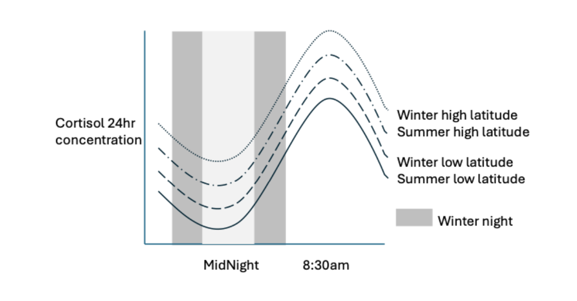
Figure 5. Circadian and seasonal rhythm of cortisol. Cortisol has a circadian rhythm and a circannual rhythm which varies with latitude. Morning cortisol levels are higher in winter and at higher latitudes.
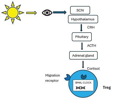
Figure 6. HPA Axis, activation of Treg intracellular circadian genes, and migration receptors. Visible sunlight stimulates the Supra-chiasmic Nucleus which in turn stimulates the hypothalamus to secrete corticotropin-releasing hormone which acts on the pituitary gland to release adrenocorticotropic hormone, which stimulates the adrenal gland to release cortisol. Cortisol interacts with its receptors which in turn activate internal circadian genes such as ‘CLOCK’ and ‘BMAL’ which control the expression of migration receptors. Migration receptors interact with chemokines to allow the egress and return migration of Tregs.
Control of Circadian Treg Circulation via Migration Receptors
Cortisol influences the migration of Tregs via their expression of chemokine receptors CCR7 and L-selectin (CD62L) [52–54] which facilitate homing to locations where the corresponding chemicals such as CCL21 are being produced [55–57].
Cortisol and glucocorticoid receptor activation influences Treg migration to the bone marrow via the expression of the receptor CXCR4 [58]. Mineralo-corticoid receptor signaling (which binds aldosterone) reduces numbers of circulating human naïve T cells and increases their CD62L, CCR7, and CXCR4 expression, [45,55–57]. CCR6 also plays a role in Treg migration to sites of an immune response [59].
To egress out of the lymph nodes, CCR7 and L-selectin expression is reduced, whilst the sphingosine-1-phosphate (S1P) receptor R1 responds to S1P, allowing extravasation into the lymph and blood [60]. Overall, there are multiple receptors and avenues for Tregs to ‘home’ to various locations, with this being mostly driven by cortisol levels activating internal cellular clock genes with the resulting circadian expression of chemokines and their receptors.
Production of New Tregs from the Thymus is Dependent on Treg Egress and Migration (via Circadian Light Pattern) via a Feedback Mechanism
Circulation of Tregs out of the thymus and into the blood circulation is closely linked to the production of naïve Tregs in the thymus in that greater circulation of Tregs out of the thymus enables the production of Tregs. Decreased migration of Tregs out of the thymus results Tregs residing for longer in the thymus resulting a negative feedback loop whereby Treg production is slowed down, and fewer new Tregs exiting the thymus [61]. From this we can surmise that the retention of Tregs in the thymus and lower production of nascent Tregs is probably occurring more in winter due to the reduced circulation associated with short day lengths. The level of T-cell receptor DNA excision circles (TREC) in blood, which are produced during the construction of the variant T-cell receptors, has been used as a method to determine the level of production of naïve T-cells [62]. Evidence using this technique suggests that more Tregs are produced in newborns after summer [37].
Maintenance of Tregs is Dependent on Circulation and Interaction with Dendritic Cells
Tregs constitute up to 50 billion cells of the T-cell compartment [63]. A reduction of 50% of cells during the daytime in winter, as observed in European studies (see above) could amount to at least 20 billion fewer cells in circulation. This has implications in terms of Tregs exposure to antigen, transition to memory cells, division and maintenance. Tregs are in a constant state of turnover, and turnover is dependent on interaction with dendritic cells, which also demonstrate a circadian pattern. It is estimated that up to 5000 T-cells can interact with one dendritic cell per hour [64] reflecting the high number of interactions that T-cells perform to maintain immune homeostasis. Dendritic cells are highly necessary to maintain peripheral Treg populations, as Treg numbers and function depend on constant recognition of the vast array of cognate autoantigens [65]. Therefore, reduced circulation of Tregs in winter results in fewer interactions with dendritic cells leading to fewer functional Tregs.
Evidence of the Effect of Photoperiod on Immune Circulation and Response from Animal Studies
Animal studies provide evidence that the effect of reduced daylight on the levels of circulating immune cells is due to visible light alone and not ultraviolet light. A study of the photoperiod on the levels of circulating T-cells in pigs, whereby UV was eliminated, demonstrated a reduction in circulating cells in the shorter daylengths, as occurs in winter [66]. Multiple studies examining the difference in immune response according to photoperiod demonstrated higher responses in winter, presumably due to decreased suppression of the immune system [67–71]. The involvement of circannual hormones other than cortisol on the number and efficacy of the immune system has also been elucidated [72,73].
Duration and Intensity of Light is Important
The finding of seasonal daylength differences in Treg numbers and function is primarily linked to the length of the daylight period causing the downstream activation of cellular clocks and migration of Tregs, via the HPA axis. However, duration of light is not the only factor. The intensity of visible light spectrum also influences the circadian rhythm. Higher intensity of light, for example over 10,000 lumens, has a greater effect on the circadian rhythm [74] and on cortisol levels [75] than lower levels. In the natural environment, exposure to bright sunny days, which have an intensity of over 100,000 lux, results in a stronger circadian rhythm which in turn has greater effect on the circulation of immune cells. Bright days usually have a higher UV index, so immune effects that are thought to be due to UV may in fact be due to the effects of brighter light on the circadian circulation of immune cells. The intensity of light certainly varies with the season. Not only are winter days short, but they are grey with lower intensity of light and lower stimulation of the SCN, and a reduced cascade of mediators resulting in a weaker activation of the cellular clock, and subsequent reduced migration of T-cells. Similarly, the brightness, or number of lumens in an indoor office or home is around 400 lux which is not conducive to a healthy circadian rhythm and immune profile. Indeed, the increase in autoimmunity and allergy prevalence at all latitudes is suggested to be associated with reduced time spent outdoors. The lack of adequate light intensity exposure would explain this in part.
Model of Decreased Duration and Intensity of Sunlight Resulting in Fewer Tregs
The sections above describe evidence supporting how seasonal variation in duration and intensity of sunlight affect circadian hormones which in turn result in in altered expression of cellular clock genes, reduced migration receptors, reduced circulating Tregs and reduced production of naïve Tregs. Fewer Tregs in circulation at the critical age of Treg ‘training’ would result in reduced exposure of Tregs to antigen or allergen, fewer memory Tregs, and overall fewer Tregs to intervene and suppress excessive immune reactions. In children living in high latitudes, with short winter day lengths, reduced levels of circulating naive T-regs over multiple winters results in fewer Tregs being exposed to antigen and trained, and fewer memory T-regs overall, potentially resulting in reduced suppressive capacity.
Seasonal Daylight is the Most Significant Factor
To date, there has not been a plausible explanation of how living in a high latitude during childhood primes the immune system to be less tolerant. Lower levels of regulatory T-cells have been associated with predisposition to autoimmunity and allergy. This paper has provided a comprehensive explanation of how short and long photoperiods, and light intensity, affect circadian factors which in turn control the number of Tregs in circulation. The visible light spectrum is the most likely factor that is influencing Treg numbers in utero and early life and is a major environmental factor in explaining the latitudinal difference in disease prevalence. The immune system is incredibly complex and there are multiple factors at play in developing predisposition to disease. Ultraviolet radiation of the skin has an effect on inflammatory mediators however it is yet to be established how cumulative years of reduced UV exposure influences the level and function of memory regulatory and effector T-cells and other disease related immune factors. Growing up in a high latitude with reduced winter daylengths and light intensity affects Treg circulation and production and the development of a tolerant immune system, thereby contributing to the predisposition to autoimmunity and allergy.
References
2. Clark MS. “I see the light”: The role of seasonal photo period in the development of immune regulation, a potential explanation for the latitude gradient of autoimmunity and allergy. Archives of Autoimmune Diseases. 2025;5(1):1–6.
3. Simpson S Jr, Wang W, Otahal P, Blizzard L, van der Mei IAF, Taylor BV. Latitude continues to be significantly associated with the prevalence of multiple sclerosis: an updated meta-analysis. J Neurol Neurosurg Psychiatry. 2019 Nov;90(11):1193–200.
4. Sabel CE, Pearson JF, Mason DF, Willoughby E, Abernethy DA, Taylor BV. The latitude gradient for multiple sclerosis prevalence is established in the early life course. Brain. 2021 Aug 17;144(7):2038–46.
5. Osborne NJ, Ukoumunne OC, Wake M, Allen KJ. Prevalence of eczema and food allergy is associated with latitude in Australia. J Allergy Clin Immunol. 2012 Mar;129(3):865–7.
6. Mullins RJ, Clark S, Camargo CA Jr. Regional variation in epinephrine autoinjector prescriptions in Australia: more evidence for the vitamin D-anaphylaxis hypothesis. Ann Allergy Asthma Immunol. 2009 Dec;103(6):488–95.
7. Hoyos-Bachiloglu R, Morales PS, Cerda J, Talesnik E, González G, Camargo CA Jr, et al. Higher latitude and lower solar radiation influence on anaphylaxis in Chilean children. Pediatr Allergy Immunol. 2014 Jun;25(4):338–43.
8. Khalili H, Huang ES, Ananthakrishnan AN, Higuchi L, Richter JM, Fuchs CS, et al. Geographical variation and incidence of inflammatory bowel disease among US women. Gut. 2012 Dec;61(12):1686–92.
9. Arkema EV, Hart JE, Bertrand KA, Laden F, Grodstein F, Rosner BA, et al. Exposure to ultraviolet-B and risk of developing rheumatoid arthritis among women in the Nurses' Health Study. Ann Rheum Dis. 2013 Apr;72(4):506–11.
10. Karvonen M, Tuomilehto J, Libman I, LaPorte R. A review of the recent epidemiological data on the worldwide incidence of type 1 (insulin-dependent) diabetes mellitus. World Health Organization DIAMOND Project Group. Diabetologia. 1993 Oct;36(10):883–92.
11. Psaroulaki E, Katsaras GN, Samartzi P, Chatziravdeli V, Psaroulaki D, Oikonomou E, et al. Association of food allergy in children with vitamin D insufficiency: a systematic review and meta-analysis. Eur J Pediatr. 2023 Apr;182(4):1533–54.
12. Falk S, Krämer J, Leson S, Pilo D, Deffner M, Ostkamp P, et al. Single-cell analysis of skin and blood reveals systemic immune responses to ultraviolet B and their impairment in MS. J Neuroinflammation. 2026 Jun 3;23(1):210.
13. Riedmann U, Dibben C, de Gruijl FR, Gorman S, Hart PH, Hoel DG, et al. Beneficial health effects of ultraviolet radiation: expert review and conference report. Photochem Photobiol Sci. 2025 Jun;24(6):867–93.
14. Johnson-Huang LM, Suárez-Fariñas M, Sullivan-Whalen M, Gilleaudeau P, Krueger JG, Lowes MA. Effective narrow-band UVB radiation therapy suppresses the IL-23/IL-17 axis in normalized psoriasis plaques. J Invest Dermatol. 2010 Nov;130(11):2654–63.
15. Clark MS, Christie M, Jones M, Ashley S, Tang MLK. Seasonal variation in sunlight exposure is differently associated with changes in T regulatory and T-helper 17 cell blood counts in adolescent and adults females: a pilot study. Photochem Photobiol Sci. 2025 Jan;24(1):23–35.
16. Hart PH, Norval M. More Than Effects in Skin: Ultraviolet Radiation-Induced Changes in Immune Cells in Human Blood. Front Immunol. 2021 Jun 10;12:694086.
17. Honing DY, Luiten RM, Matos TR. Regulatory T Cell Dysfunction in Autoimmune Diseases. Int J Mol Sci. 2024 Jun 29;25(13):7171.
18. Mexhitaj I, Nyirenda MH, Li R, O'Mahony J, Rezk A, Rozenberg A, et al. Abnormal effector and regulatory T cell subsets in paediatric-onset multiple sclerosis. Brain. 2019 Mar 1;142(3):617–32.
19. Verma ND, Lam AD, Chiu C, Tran GT, Hall BM, Hodgkinson SJ. Multiple sclerosis patients have reduced resting and increased activated CD4+CD25+FOXP3+T regulatory cells. Sci Rep. 2021 May 18;11(1):10476.
20. Bossowski A, Moniuszko M, Idźkowska E, Grubczak K, Singh P, Bossowska A, et al. Decreased proportions of CD4 + IL17+/CD4 + CD25 + CD127− and CD4 + IL17+/CD4 + CD25 + CD127 − FoxP3+ T cells in children with autoimmune thyroid diseases*. Autoimmunity. 2016 Aug;49(5):320–8.
21. Prince BT, Devonshire AL, Erickson KA, Bergerson J, Fuleihan D, Szychlinski C, et al. Regulatory T-cell populations in children are affected by age and food allergy diagnosis. J Allergy Clin Immunol. 2017 Oct;140(4):1194–6.e16.
22. Palomares O. The role of regulatory T cells in IgE-mediated food allergy. J Investig Allergol Clin Immunol. 2013;23(6):371–82
23. Turner JA, Stephen-Victor E, Wang S, Rivas MN, Abdel-Gadir A, Harb H, et al. Regulatory T cell-derived TGF-β1 controls multiple checkpoints governing allergy and autoimmunity. Immunity. 2020 Dec 15;53(6):1202–14.
24. Rivas MN, Chatila TA. Regulatory T cells in allergic diseases. Journal of Allergy and Clinical Immunology. 2016 Sep 1;138(3):639–52.
25. Tulic MK, Andrews D, Crook ML, Charles A, Tourigny MR, Moqbel R, et al. Changes in thymic regulatory T-cell maturation from birth to puberty: differences in atopic children. J Allergy Clin Immunol. 2012 Jan;129(1):199–206.e1–4.
26. Quilis N, Mesa-Del-Castillo Bermejo P, Boix P, Juanola O, Bernabeu P, Francés R, et al. Peripheral blood regulatory T cells and disease activity, quality of life, and outcomes in children with juvenile idiopathic arthritis. Pediatr Rheumatol Online J. 2024 Aug 1;22(1):69.
27. Wang L, Liang Y, Zhao C, Ma P, Zeng S, Ju D, et al. Regulatory T cells in homeostasis and disease: molecular mechanisms and therapeutic potential. Signal Transduct Target Ther. 2025 Oct 14;10(1):345.
28. Godfrey VL, Wilkinson JE, Russell LB. X-linked lymphoreticular disease in the scurfy (sf) mutant mouse. Am J Pathol. 1991 Jun;138(6):1379–87.
29. Darrasse-Jèze G, Marodon G, Salomon BL, Catala M, Klatzmann D. Ontogeny of CD4+CD25+ regulatory/suppressor T cells in human fetuses. Blood. 2005 Jun 15;105(12):4715–21.
30. Rocamora-Reverte L, Melzer FL, Würzner R, Weinberger B. The Complex Role of Regulatory T Cells in Immunity and Aging. Front Immunol. 2021 Jan 27;11:616949.
31. Santner-Nanan B, Seddiki N, Zhu E, Quent V, Kelleher A, Fazekas de St Groth B, et al. Accelerated age-dependent transition of human regulatory T cells to effector memory phenotype. Int Immunol. 2008 Mar;20(3):375–83.
32. Hayakawa S, Ohno N, Okada S, Kobayashi M. Significant augmentation of regulatory T cell numbers occurs during the early neonatal period. Clin Exp Immunol. 2017 Nov;190(2):268–79.
33. Singh VR, O'Donnell LA. Age-Stratified Treg Responses During Viral Infections of the Central Nervous System: A Literature Review. J Med Virol. 2025 Apr;97(4):e70315.
34. Srinivasan J, Lancaster JN, Singarapu N, Hale LP, Ehrlich LIR, Richie ER. Age-Related Changes in Thymic Central Tolerance. Front Immunol. 2021 Apr 22;12:676236.
35. van Gent R, van Tilburg CM, Nibbelke EE, Otto SA, Gaiser JF, Janssens-Korpela PL, et al. Refined characterization and reference values of the pediatric T- and B-cell compartments. Clin Immunol. 2009 Oct;133(1):95–107.
36. Luo Y, Acevedo D, Vlagea A, Codina A, García-García A, Deyà-Martínez A, et al. Changes in Treg and Breg cells in a healthy pediatric population. Front Immunol. 2023 Nov 21;14:1283981
37. Disanto G, Watson CT, Meier UC, Ebers GC, Giovannoni G, Ramagopalan SV. Month of birth and thymic output. JAMA Neurol. 2013 Apr;70(4):527–8.
38. Ramos-Leví AM, Collado G, Marazuela M. Seasonality of month of birth in patients with autoimmune endocrine diseases: A systematic review. Endocrinol Diabetes Nutr (Engl Ed). 2022 Dec;69(10):779–90.
39. Lange T, Luebber F, Grasshoff H, Besedovsky L. The contribution of sleep to the neuroendocrine regulation of rhythms in human leukocyte traffic. Semin Immunopathol. 2022 Mar;44(2):239–54.
40. Lamikanra AA, Tsang HP, Elsiddig S, Spencer M, Curnow E, Danby R, et al. The Migratory Properties and Numbers of T Regulatory Cell Subsets in Circulation Are Differentially Influenced by Season and Are Associated With Vitamin D Status. Front Immunol. 2020 May 19;11:685.
41. Maboshe W, Macdonald HM, Wassall H, Fraser WD, Tang JCY, Fielding S, et al. Low-Dose Vitamin D3 Supplementation Does Not Affect Natural Regulatory T Cell Population but Attenuates Seasonal Changes in T Cell-Produced IFN-γ: Results From the D-SIRe2 Randomized Controlled Trial. Front Immunol. 2021 Jun 28;12:623087.
42. Schweintzger NA, Gruber-Wackernagel A, Shirsath N, Quehenberger F, Obermayer-Pietsch B, Wolf P. Influence of the season on vitamin D levels and regulatory T cells in patients with polymorphic light eruption. Photochem Photobiol Sci. 2016 Mar;15(3):440–6.
43. Dimitrov S, Benedict C, Heutling D, Westermann J, Born J, Lange T. Cortisol and epinephrine control opposing circadian rhythms in T cell subsets. Blood. 2009 May 21;113(21):5134–43.
44. Bollinger T, Bollinger A, Skrum L, Dimitrov S, Lange T, Solbach W. Sleep-dependent activity of T cells and regulatory T cells. Clin Exp Immunol. 2009 Feb;155(2):231–8.
45. Besedovsky L, Dimitrov S, Born J, Lange T. Nocturnal sleep uniformly reduces numbers of different T-cell subsets in the blood of healthy men. Am J Physiol Regul Integr Comp Physiol. 2016 Oct 1;311(4):R637–42.
46. Cain DW, Cidlowski JA. Immune regulation by glucocorticoids. Nat Rev Immunol. 2017 Apr;17(4):233–47.
47. Hadlow N, Brown S, Wardrop R, Conradie J, Henley D. Where in the world? Latitude, longitude and season contribute to the complex co-ordinates determining cortisol levels. Clin Endocrinol (Oxf). 2018 Sep;89(3):299–307.
48. Tendler A, Bar A, Mendelsohn-Cohen N, Karin O, Korem Kohanim Y, Maimon L, et al. Hormone seasonality in medical records suggests circannual endocrine circuits. Proc Natl Acad Sci U S A. 2021 Feb 16;118(7):e2003926118.
49. He W, Holtkamp S, Hergenhan SM, Kraus K, de Juan A, Weber J, et al. Circadian Expression of Migratory Factors Establishes Lineage-Specific Signatures that Guide the Homing of Leukocyte Subsets to Tissues. Immunity. 2018 Dec 18;49(6):1175–90.
50. Szredzka A, Chwastowicz A, Pergoł J, Matryba P. Circadian clocks and adaptive immune function: from mechanisms to therapeutic applications. Front Immunol. 2025 Dec 3;16:1697854.
51. Yu X, Rollins D, Ruhn KA, Stubblefield JJ, Green CB, Kashiwada M, et al. TH17 cell differentiation is regulated by the circadian clock. Science. 2013 Nov 8;342(6159):727–30.
52. Venturi GM, Conway RM, Steeber DA, Tedder TF. CD25+CD4+ regulatory T cell migration requires L-selectin expression: L-selectin transcriptional regulation balances constitutive receptor turnover. J Immunol. 2007 Jan 1;178(1):291–300.
53. Lim HW, Hillsamer P, Kim CH. Regulatory T cells can migrate to follicles upon T cell activation and suppress GC-Th cells and GC-Th cell-driven B cell responses. J Clin Invest. 2004 Dec;114(11):1640–9.
54. Ochando JC, Yopp AC, Yang Y, Garin A, Li Y, Boros P, et al. Lymph node occupancy is required for the peripheral development of alloantigen-specific Foxp3+ regulatory T cells. J Immunol. 2005 Jun 1;174(11):6993–7005.
55. Besedovsky L, Born J, Lange T. Endogenous glucocorticoid receptor signaling drives rhythmic changes in human T-cell subset numbers and the expression of the chemokine receptor CXCR4. FASEB J. 2014 Jan;28(1):67–75.
56. Besedovsky L, Linz B, Born J, Lange T. Mineralocorticoid receptor signaling reduces numbers of circulating human naïve T cells and increases their CD62L, CCR7, and CXCR4 expression. Eur J Immunol. 2014 Jun;44(6):1759–69.
57. Besedovsky L, Linz B, Dimitrov S, Groch S, Born J, Lange T. Cortisol increases CXCR4 expression but does not affect CD62L and CCR7 levels on specific T cell subsets in humans. Am J Physiol Endocrinol Metab. 2014 Jun 1;306(11):E1322–9.
58. Yuan Y, Wu S, Li W, He W. A Tissue-Specific Rhythmic Recruitment Pattern of Leukocyte Subsets. Front Immunol. 2020 Feb 14;11:102.
59. Cowan JE, Baik S, McCarthy NI, Parnell SM, White AJ, Jenkinson WE, et al. Aire controls the recirculation of murine Foxp3+ regulatory T-cells back to the thymus. Eur J Immunol. 2018 May;48(5):844–54.
60. Garcia-Seyda N, Song S, Seveau de Noray V, David-Broglio L, Matti C, Artinger M, et al. Naive T lymphocytes chemotax long distance to CCL21 but not to a source of bioactive S1P. iScience. 2023 Aug 19;26(10):107695.
61. Thiault N, Darrigues J, Adoue V, Gros M, Binet B, Perals C, et al. Peripheral regulatory T lymphocytes recirculating to the thymus suppress the development of their precursors. Nat Immunol. 2015 Jun;16(6):628–34.
62. Haas J, Würthwein C, Korporal-Kuhnke M, Viehoever A, Jarius S, Ruck T, et al. Alemtuzumab in Multiple Sclerosis: Short- and Long-Term Effects of Immunodepletion on the Peripheral Treg Compartment. Front Immunol. 2019 Jun 4;10:1204.
63. Horwitz DA, Fahmy TM, Piccirillo CA, La Cava A. Rebalancing Immune Homeostasis to Treat Autoimmune Diseases. Trends Immunol. 2019 Oct;40(10):888–908.
64. Garbi N, Hämmerling GJ, Probst HC, van den Broek M. Tonic T cell signalling and T cell tolerance as opposite effects of self-recognition on dendritic cells. Curr Opin Immunol. 2010 Oct;22(5):601–8.
65. Schildknecht A, Brauer S, Brenner C, Lahl K, Schild H, Sparwasser T, et al. FoxP3+ regulatory T cells essentially contribute to peripheral CD8+ T-cell tolerance induced by steady-state dendritic cells. Proc Natl Acad Sci U S A. 2010 Jan 5;107(1):199–203.
66. Engert LC, Weiler U, Pfaffinger B, Stefanski V, Schmucker SS. Photoperiodic Effects on Diurnal Rhythms in Cell Numbers of Peripheral Leukocytes in Domestic Pigs. Front Immunol. 2019 Mar 12;10:393.
67. Prendergast BJ, Baillie SR, Dhabhar FS. Gonadal hormone-dependent and -independent regulation of immune function by photoperiod in Siberian hamsters. Am J Physiol Regul Integr Comp Physiol. 2008 Feb;294(2):R384–92.
68. Prendergast BJ, Pyter LM. Photoperiod history differentially impacts reproduction and immune function in adult Siberian hamsters. J Biol Rhythms. 2009 Dec;24(6):509–22.
69. Prendergast BJ, Kampf-Lassin A, Yee JR, Galang J, McMaster N, Kay LM. Winter day lengths enhance T lymphocyte phenotypes, inhibit cytokine responses, and attenuate behavioral symptoms of infection in laboratory rats. Brain Behav Immun. 2007 Nov;21(8):1096–108.
70. Onishi KG, Maneval AC, Cable EC, Tuohy MC, Scasny AJ, et al. Circadian and circannual timescales interact to generate seasonal changes in immune function. Brain Behav Immun. 2020 Jan;83:33–43.
71. Stevenson TJ, Prendergast BJ. Photoperiodic time measurement and seasonal immunological plasticity. Front Neuroendocrinol. 2015 Apr;37:76–88.
72. Walton JC, Weil ZM, Nelson RJ. Influence of photoperiod on hormones, behavior, and immune function. Front Neuroendocrinol. 2011 Aug;32(3):303–19.
73. Weil ZM, Borniger JC, Cisse YM, Abi Salloum BA, Nelson RJ. Neuroendocrine control of photoperiodic changes in immune function. Front Neuroendocrinol. 2015 Apr;37:108–18.
74. Levitan RD. What is the optimal implementation of bright light therapy for seasonal affective disorder (SAD)? J Psychiatry Neurosci. 2005 Jan;30(1):72.
75. Jung CM, Khalsa SB, Scheer FA, Cajochen C, Lockley SW, Czeisler CA, et al. Acute effects of bright light exposure on cortisol levels. J Biol Rhythms. 2010 Jun;25(3):208–16.