

Abstract
The interactions between the DIX domains of three proteins (Axin1/2, Dishevelled1/2/3, and Coiled-coil-DIX1) are essential to the mechanism of action downstream of the Wnt/β-catenin signaling pathway. Structural and biophysical studies have shown that DIX domains polymerize via head-to-tail interface interactions. In our recently published study entitled 'Exploring DIX-DIX Homo- and Hetero-Oligomers in Wnt Signaling with AlphaFold2", we examined the monomer structures of DIX domains, and DIX-mediated homodimers and heterodimers with AlphaFold2 (AF2) ColabFold. First, we evaluated the AF2-based prediction by comparing the reported monomer and complex structures of the DIX domains. The results showed that AF2 is an excellent tool for predicting the 3D structures of monomers of DIX domains and of homodimers and heterodimers of DIX-mediated proteins. Second, we evaluated the calculated binding affinities (KD) of DIX domains using the PRODIGY method. The results showed that the calculated KD values are compatible with experimentally obtained values. In this commentary, we present new computational results using AlphaFold3 (AF3), the latest version of AlphaFold, and VD-MM/GBSA calculations with HawkDock2. Overall, AF2/3 and other bioinformatics tools provide valuable insights into the DIX-mediated signaling pathway at the molecular level.
Keywords
AlphaFold2/3, HawkDock2, VD-MM/GBSA, DIX domain, Protein-Protein Interactions, Wnt signaling
Background on the Role of DIX-Mediated Interactions
Wnt signaling pathways are fundamental to the regulation of cell proliferation and differentiation, and their dysregulation is implicated in various pathologies, including cancer [1,2]. The Wnt/β-catenin signaling is initiated and regulated by protein-protein interactions, such as Wnt and Frizzled [3], Frizzled and Dishevelled [4] and Dishevelled and Axin [5,6]. Investigating these protein-protein interactions (PPIs) has therefore been of significant interest for understanding the mechanism of action of the Wnt/β-catenin signaling. One of the PPIs is DIX-mediated interactions. In Wnt/β-catenin signaling, three DIX proteins, Axin, Dishevelled (Dvls), and Coiled-coil-DIX1 (Ccd1), are central components [7–11]. Through their DIX domains (Figure 1A), these proteins can form dynamic homo- or heteropolymers in vitro and in vivo, leading to either the autoinhibition or the activation of downstream Wnt signaling [7,10,12,13]. However, the precise mechanisms of DIX-mediated homomeric and heteromeric polymerization remain unclear. Moreover, whether the roles of their paralogs (Axin1/2, Dvl1/2/3, and Ccd1) are complementary or redundant remains unresolved [14,15].
DIX domains adopt a ubiquitin-like fold structure, characterized by five b-strands (b1-b5) and one a-helix (a1) (Figure 1A) [6,10,16,17]. These domains form dimers or oligomers through head-to-tail interactions. For example, the DAX1-DAX1 homodimer forms a head-to-tail complex, showing that the tail (b4) of DAX1 interacts with the head (b2) of DAX1. High-resolution data from X-ray crystallography, Nuclear Magnetic Resonance (NMR) spectroscopy, and site-directed mutagenesis have successfully identified the critical residues, such as Y796, V836, and F837, in human Axin1, driving this dimerization [12,13,16,18]. Despite detailed structural insights, quantifying the binding affinities of DIX-containing proteins remains challenging due to technical limitations. Since DIX domains can form dimers or oligomers even at very low concentrations, purifying the monomeric form of the DIX domain is difficult yet essential for accurately determining homodimer and heterodimeric binding affinities. To overcome this challenge, many studies have employed mutants to estimate the binding affinities of DIX-domain homodimers and heterodimers [8,10]. However, DIX mutants can still form oligomers or dimers at low concentration, and the stability of the mutant proteins remains uncertain [13].
To explore the binding and specificity of DIX domain-mediated interactions, we used AF2 and the PRODIGY (PROtein binDIng enerGY prediction) contact-based prediction approach. For comparison, we also used the HawkDock server(ver 1) to estimate the relative binding affinities of DIX domains using the MM-GBSA method [19,20]. In our recently published work, the results strongly support the conclusion that AF2 ColabFold (ver 1.6.1) is an easy-to-use, fast tool that, remarkably, can accurately generate 3D structures of the DIX domain and its complexes [21–24]. Our results partially explain how DIX-mediated interactions among these three proteins and their paralogs may exhibit distinct mechanisms of action in the Wnt/β-catenin signaling.
Findings of the Published Work
In our previous work, we used two approaches to predict the structures of DIX domain complexes. The first approach utilized the AF2 ColabFold with a residue index gap of 200 between chains, such as "DAX1(Axin1 DIX domain): DAX1(Axin1 DIX domain)" [23]. The 1:1 binding mode was used. The second approach involved introducing the extra amino acid residues between two target proteins. Eighteen residues (six GGS repeats) were used to link two target domains. For example, we predicted the complex structure of DAX1 homodimer using the DAX1-(GGS)6-DAX1 construct. For the DAX1(Axin1 DIX):DIX2 (Dvl2 DIX) heterodimer, we used the sequence of DAX1-(GGS)6-DIX2 and DIX2-(GGS)6-DAX1. Remarkably, the DAX1-(GGS)6-DIX2 complex structure revealed an interaction between the tail interface of DAX1 and the head interface of DIX2. The DIX2-(GGS)6-DAX1 complex structure revealed interactions between the tail interface of DIX2 and the head interface of DAX1 (Figure S1). All predicted homodimers and heterodimers share similar structural features; however, the binding regions (especially the head interfaces) of the complexes are relatively flexible [24]. In addition, the complex structures of homodimers and heterodimers support the previous conclusion that DIX-mediated homotypic and heterotypic interactions share the same head and tail residues [12]. For example, previous studies reported that Y27 of human DVL2 DIX (DIX2), corresponding to Y796 of human Axin1 DIX (DAX1), is a critical residue forming the homo- and heterodimer. We used the nomenclature of mutants M2, M3, and M4 based on Fielder et al.'s work [12] (Figure 1A). These mutations are located outside the complex interfaces of the DIX domains and are therefore not expected to affect protein-protein interactions. AF2 generated five models (rank 1-5) for each complex. The pLDDT value was used to rank the predicted models by confidence level [21,25–27]. The top-ranked (rank 1) complex model for each DIX homodimer and heterodimer was selected for further analysis. The 6xGGS linker was removed from the AF2-predicted complex structures prior to further analysis.
We found that AF2 accurately predicted the 3D structures of DIX domains [24]. The predicted monomer DIX structures superimpose well onto the previously reported X-ray structures of DIX proteins, with an overall root-mean-square deviation (RMSD) of less than 0.5 Å [24]. We then generated the homodimer of Dvl2 DIX-M2(Y27W) mutant and heterodimer of wild-type Axin1 DIX (DAX1) and Dvl1 DIX(DIX1) to evaluate the performance of AF2 by comparison with the corresponding reported X-ray structures [18]. Both DIX-domain complexes exhibited head-to-tail interactions, consistent with prior reports. The backbone RMSD between predicted and X-ray structures is also less than 0.5 Å, supporting that AF2 can predict DIX-DIX complex structures with great accuracy. The orientation of the side chains of amino acid residues in the predicted homodimer and heterodimer is remarkably similar to that observed in experimental structures. For example, AF2 predicted that the side chain of E23 forms a hydrogen bond with the side chain of W27 of DIX-M2 and a salt bridge with the side chain of K68 of the adjacent molecule, as found in the DIX-M2(Y27W) structure [18]. We further evaluated the AF2-prediction complex using the DIX mutants, DIX2-M4(Y27D)-(GGS)6-DAX1-M2(V836A/F837A) and DAX1-M2-(GGS)6-DIX2-M4 mutants. DIX2-M4(Y27D) is a mutant in the head interface. DAX2-M2(V836A/F837A) is a mutant in the tail interface [12]. Since the mutations are located outside the binding interfaces, we expected that DIX2-M4-(GGS)6-DAX1-M2 would form the complex via the tail interface of DIX2-M4 and the head interface of DAX1-M2. Indeed, AF2 predicted the complex structure of DIX2-M4(Y27D)-(GGS)6-DAX1-M2(V836A/F837A) [23]. As a negative control, the complex structure of DAX1-M2(V836A/F837A)-(GGS)6-DIX2-M4(Y27D) was also generated to see whether it might predict the head-to-tail interaction. Interestingly, the predicted complex of DAX1-M2-(GGS)6-DIX2-M4 showed diverse structures. Two structures are the same as that of DIX2-M4(Y27D)-(GGS)6-DAX1-M2(V836A/F837A). One of the predicted complexes showed no interaction between the two DIX mutants [7]. The results suggest that AlphaFold2-powered ColabFold is an excellent method for predicting the 3D structures of homodimers and heterodimers of DIX-containing proteins. The results imply that the AF2 may be used to investigate the effects of mutations on protein-protein interactions [25].
The PRODIGY server was used to calculate the binding affinities of the DIX complexes. The results show that the binding affinity (KD) of the Axin2 DIX(DAX2) homodimer is stronger than that of the Axin1 DIX(DAX1) homodimer. For Dishevelled (Dvl) proteins, Dvl1 DIX homodimer (DIX1-DIX1) shows higher binding affinity than Dvl2 DIX(DIX2) and Dvl3 DIX(DIX3) homodimers. For the positive Wnt signaling regulator Coiled-coil-DIX1(Ccd1), the KD value of the Ccd1 DIX homodimer is lower than that of the Axin1 DIX(DAX1) homodimer. The relative binding strength order of homodimers was predicted to be DAX2 > DC1 > DIX1 >> DIX2 ~ DIX3. The weaker interactions of Dvl2/3 DIX may explain the dynamics of Dvl oligomerization [7,12]. The differing binding affinities between Axin1/2 DIX domains and Dvl1 versus Dvl2/3 DIX domains suggest that these protein paralogs may have distinct roles in Wnt signaling [14,15]. The strong DAX-DAX homotypic interaction may explain why cytoplasmic Dvl is unable to disrupt DAX-DAX interactions [16].
For heterodimers, we found that their binding affinities are predicted to be stronger than DIX2/3 homodimers but weaker than Axin1/2 DIX homodimers. Remarkably, the binding affinity of Dvl2/3 DIX(DIX2/3)↔Axin1 DIX(DAX1) interaction is 10x ~ 70x weaker than Axin1 DIX(DAX1)↔Dvl2/3 DIX(DIX2/3) interaction [24]. The predicted results support a study reporting that DAX1 might control the oligomerization of the DIX2 [16]. Kan et al. found that DAX1 binds to the ends of Dvl oligomers, suggesting that roughly matched numbers of Axin and Dvl are associated with the activated receptors [16]. The overall results imply that the difference in binding affinity between DAX-DIX and DIX-DAX controls the size of Dvl oligomers.
For comparison, the HawkDock server was used to calculate binding energies for the DIX domain via molecular docking and the molecular mechanics/generalized MM/GBSA method [19]. However, the binding affinities of DIX-mediated interactions obtained by the MM/GBSA method were quite higher than the reported experimental values (Table 1). In addition, employing HawkDock to assess the binding affinities of the predicted DIX homodimers and heterodimers required the preliminary separation of AF2-predicted complexes into monomers, followed by an independent redocking procedure at the HawkDock server. The docking structures of DIXs derived from the predicted monomer structures were not as accurate as the AF2-predicted complex structures.
|
Complex |
aΔG (kcal/mol) |
aΔG (kcal/mol) |
cΔG (kcal/mol) |
Exp (KD, mM) |
|
|
AF2 > VD-MM/GBSA |
AF3 > VD-MM/GBSA |
AF2 > PRODIGY |
|
|
DAX1↔DAX1 |
–116.86 |
–103.95 |
–8.4 |
d 0.24 |
|
DIX2↔DIX2 |
–52.94 |
–74.49 |
–7.5 |
e 5-20 |
|
DIX2↔DAX1 |
–91.10 |
–93.81 |
–8.2 |
d 9 |
|
DAX1↔DIX2 |
–111.61 |
b –81.92 |
–8.5 |
d 24 |
|
a This work b Third top-ranked complex (rank 3) of DAX1-DIX2 was used to calculate ΔG c Ref. [24]. d Ref. [16]. The detailed information of the protein-protein interaction: DAX1↔DAX1,KD = 0.24 µM; DAX1-M3↔DIX2-M2, KD=24 µM; DIX2-M2↔DAX1-M2, KD = 9 µM e Ref. [12]. The detailed information of the protein-protein interaction: DAX1-M3↔DAX1-M2, KD = 45 µM ; DIX2↔DIX2,KD = 5–20 µM. |
||||
Comparison of AF2 and AF3; HawkDock and HawkDock2
Following the publication of AF2-based analyses of DIX domain-mediated interactions, the latest version of AlphaFold, AlphaFold3 (AF3), has been released. AF3 incorporates the Pairformer module, which replaces the Evoformer module in AF2 [27,28]. While AF2 relies heavily on the Multiple Sequence Alignment (MSA)-generated embeddings for predicting protein interactions, AF3 simplifies MSA processing by reducing the number of MSA blocks to four [28]. Consequently, AF3 addresses many of the limitations of AF2, including the prediction of protein-protein and protein-ligand interactions, as well as post-translational modifications. In parallel, HawkDock server 2 (HawkDock2) was released, which can predict binding affinities of complexes predicted by AF2/3 or other AI-driven protein prediction methods, without requiring additional structural preparation. Moreover, HawkDock2 can decompose residue-level energy contributions, especially at the interaction interfaces, using the VD-MM/GBSA method [29]. This VD-MM/GBSA method is reported to outperform conventional MM/GBSA approaches in predicting binding affinities [29].
In this commentary, we re-evaluated AF2- and AF3-predicted structures of the DAX1↔DAX1, DIX2↔DIX2, DIX2↔DAX1, and DAX1↔DIX2 complexes. Using the AF2 ColabFold server (Ver. 1.6.1), the five top-ranked models for all four systems reproduced the expected head-to-tail arrangements: DAX1↔DAX1 and DIX2↔DIX2 homodimers, the DIX2↔DAX1 heterodimer formed by the DIX2 tail and the DAX1 head, and the DAX1↔DIX2 heterodimer formed by the DAX1 tail and the DIX2 head (Figure S1). The AF3 server produced highly comparable models for the two homodimers and for the DIX2↔DAX1 heterodimer. However, the DAX1-(GGS)6-DIX2 input construct produced two different distributions of AF3 models. Three of the five top-ranked AF3 (third to fifth ranked) models retained the expected DAX1-tail/DIX2-head interaction, whereas two top-ranked (first and second ranked) models adopted an alternative DIX2-tail/DAX1-head arrangement (Figures S1B and S2). Because AF2 and AF3 use different network architectures, Evoformer and Pairformer, respectively, these results suggest that both platforms should be considered complementary when evaluating DIX-mediated heterodimer structures. One possible explanation is that AF3’s reduced dependence on deep multiple sequence alignments may affect the ranking or prioritization of alternative interaction modes when co-evolutionary constraints are weak, as may be the case for the specific DAX1-tail/DIX2-head interface.
The binding affinities estimated by the VD-MM/GBSA method were also broadly consistent between AF2- and AF3-derived complexes. Although the VD-MM/GBSA estimation is overestimated, the relative order of binding energies was DAX1↔DAX1 > DIX2↔DAX1 > DIX2↔DIX2, like our previous report [24] (Table 1).
The advantage of the VD-MM/GBSA method is that it enables dissection of the energy contribution of each residue at the interaction interfaces (Figures 1B and 1D). For DAX1-DAX1, the D862, F831, Y823 residues of the tail of DAX1, and the T815, Y796, and I794 residues of the head of DAX1 are the major residues involved in the interaction. While previous studies have focused on residue Y796 of DAX1, this work identified additional critical residues. In particular, the F831 and T815 residues of DAX1 contribute to stabilizing the homodimer. For DIX2-DIX2, the K68, V67, E69 residues of the tail of DIX2, and the L28, Y27, and R84 residues of the head of the second DIX2 stabilize the complex structure. While previous studies focused on residues Y27 of DIX2 and Y796 of DAX1 for protein-protein interactions, our analysis clearly demonstrates that additional residues at the head and tail interfaces may also play important roles in stabilizing DIX-mediated homo- and heterodimers. For DIX2-DIX2 homodimer formation, the residue R84 of DIX2 is also important. For DIX2-DAX1 interaction, R797, Y796, and I794 residues of DAX1, and F64, D61, and F56 residues of DIX2 are important contributors to the binding affinity. In the DAX1-DIX2 complex structure, F831 and Y823 of DAX1, and the Y27, L28, and T25 residues of DIX2 are the primary contributors to heterodimer stabilization. The residues involved in DIX-mediated interactions are shown in Figure 1C. Therefore, follow-up studies, including site-directed mutagenesis and quantitative binding assays, will be needed to define the functional contributions of these newly identified residues to DIX-domain-mediated protein-protein interactions. We also dissected the binding energies of the AF3-predicted complex structure (Figure 1D), which are similar to those of the AF2-predicted complex (Figure 1B).
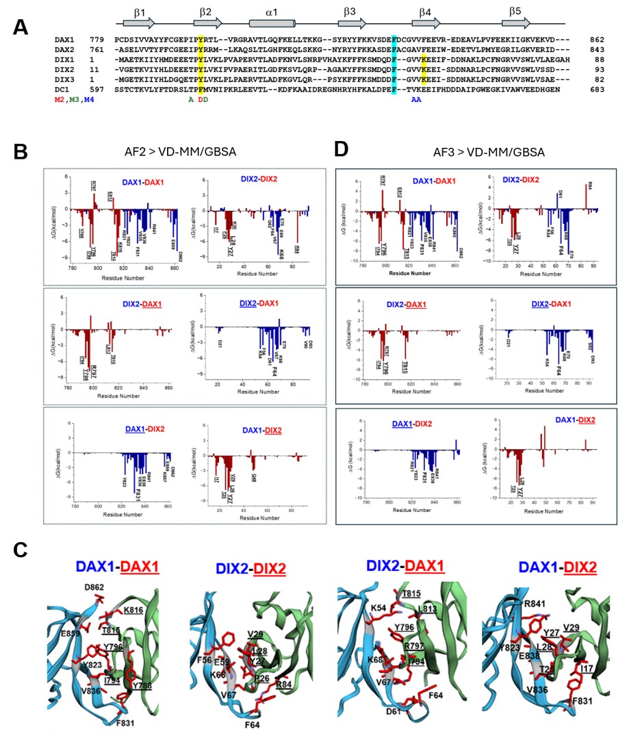
Figure 1. Structural organization and residue-level binding-energy analysis of DIX-domain complexes. (A) Sequence alignment of the DIX domains of Axin, Dishevelled (Dvl), and Coiled-coil-DIX1 (Ccd1), including their paralogs and mutants (M2, M3, and M4). The secondary-structure elements of the DIX domain are indicated, consisting of five β-strands and one α-helix. (B) VD-MM/GBSA decomposition of AF2-predicted DIX-domain complexes using HawkDock2. The DAX1↔DAX1, DIX2↔DIX2, DIX2↔DAX1, and DAX1↔DIX2 complexes were analyzed based on the top-ranked conformer (rank 1). Tail interfaces are shown in blue and head interfaces of the interacting partner are shown in red; residues that make major energetic contributions to binding are highlighted at the protein-protein interface. (C) Cartoon representations of the four AF2-predicted DIX-domain complexes, with amino acids predicted to be important for protein-protein binding shown as stick models and labeled with their residue numbers. (D) VD-MM/GBSA decomposition of AF3-predicted DIX-domain complexes. Unless otherwise indicated, AF3-based VD-MM/GBSA calculations were performed using the top-ranked conformer (rank 1). For the DAX1-tail/DIX2-head arrangement, however, the DAX1-DIX2 structure predicted as rank 3 was used for the calculation (See Figures S1 and S2).
Conclusion
Characterizing protein-protein interactions and their specificity is crucial for understanding the mechanism of action in Wnt signaling. Because of the diversity of cellular proteins and the experimental challenges associated with measuring DIX-mediated interactions, theoretical approaches such as homology modeling, docking, and molecular dynamics provide valuable complementary tools. In our previous work, we used AF2-powered ColabFold to generate homodimers and heterodimers of DIX domains without templates, demonstrating its strong performance. We also obtained binding affinities for DIX-mediated interactions using the PRODIGY server based on AF2-predicted and optimized structures. The predicted KD values for DIX-DIX interactions were broadly comparable to reported experimental data and partially explained the molecular mechanisms by which Axin1/2, Dvl1/2/3, and Ccd1 regulate intracellular Wnt signaling. In this commentary, we used AF3 to generate DAX1↔DAX1, DIX2↔DIX2, DIX2↔DAX1, and DAX1↔DIX2 complexes using the two approaches described in the previous study [24]. AF3 produced results that were generally consistent with AF2 for the DAX1↔DAX1 and DIX2↔DIX2 homodimers and for the DIX2↔DAX1 heterodimer. In contrast, AF3 showed a distinct prediction pattern for the DAX1-(GGS)6-DIX2 input construct, yielding both the expected DAX1-tail/DIX2-head arrangement and alternative DIX2-tail/DAX1-head models. This finding highlights the complementary utility of AF2 and AF3 for validating predicted DIX-domain complex structures. Using HawkDock2, we identified critical residues at the head and tail interfaces of the AF2- and AF3-predicted DIX complexes that had not been previously recognized, providing testable targets for future biological experiments. Overall, our results support the conclusion that AF2-powered ColabFold and AF3, in conjunction with HawkDock2, are easy-to-use and rapid approaches for exploring previously uncharacterized protein-protein interactions at the atomic level. Because DIX oligomerization is modulated by post-translational modifications such as ubiquitination and phosphorylation [9], AF3 may also be useful for examining how these modifications affect the structures of Dvl and Axin proteins, thereby deepening our understanding of DIX-mediated Wnt signaling.
Supporting Information
Acknowledgments
I am expressing my sincere gratitude to LeMoyne-Owen College for the vital support of this work.
Conflicts of Interest
The author declares that the research was conducted in the absence of any commercial or financial relationships that could be construed as a potential conflict of interest.
AI Disclosure
Artificial intelligence tools were used only for language editing and grammar checking. The overall manuscript, including the research design, analysis, interpretation, and scientific content, was written and approved by the author.
References
2. Park WJ, Kim MJ. A New Wave of Targeting 'Undruggable' Wnt Signaling for Cancer Therapy: Challenges and Opportunities. Cells. 2023 Apr 8;12(8):1110.
3. Hirai H, Matoba K, Mihara E, Arimori T, Takagi J. Crystal structure of a mammalian Wnt-frizzled complex. Nat Struct Mol Biol. 2019 May;26(5):372–9.
4. Qian Y, Ma Z, Xu Z, Duan Y, Xiong Y, Xia R, et al. Structural basis of Frizzled 4 in recognition of Dishevelled 2 unveils mechanism of WNT signaling activation. Nat Commun. 2024 Sep 2;15(1):7644.
5. Ehebauer MT, Arias AM. The structural and functional determinants of the Axin and Dishevelled DIX domains. BMC Struct Biol. 2009 Nov 12;9:70.
6. Yamanishi K, Kumano W, Terawaki SI, Higuchi Y, Shibata N. Head-to-Tail Complex of Dishevelled and Axin-DIX Domains: Expression, Purification, Crystallographic Studies and Packing Analysis. Protein Pept Lett. 2019;26(10):792–7.
7. Schwarz-Romond T, Fiedler M, Shibata N, Butler PJ, Kikuchi A, Higuchi Y, Bienz M. The DIX domain of Dishevelled confers Wnt signaling by dynamic polymerization. Nat Struct Mol Biol. 2007 Jun;14(6):484–92.
8. Liu YT, Dan QJ, Wang J, Feng Y, Chen L, Liang J, et al. Molecular basis of Wnt activation via the DIX domain protein Ccd1. J Biol Chem. 2011 Mar 11;286(10):8597–608.
9. Madrzak J, Fiedler M, Johnson CM, Ewan R, Knebel A, Bienz M, et al. Ubiquitination of the Dishevelled DIX domain blocks its head-to-tail polymerization. Nat Commun. 2015 Apr 24;6:6718.
10. Terawaki SI, Fujita S, Katsutani T, Shiomi K, Keino-Masu K, Masu M, et al. Structural basis for Ccd1 auto-inhibition in the Wnt pathway through homomerization of the DIX domain. Sci Rep. 2017 Aug 10;7(1):7739.
11. Wang L, Zhu R, Wen Z, Fan HS, Norwood-Jackson T, Jathan D, et al. Structural and Functional Insights into Dishevelled-Mediated Wnt Signaling. Cells. 2024 Nov 11;13(22):1870.
12. Fiedler M, Mendoza-Topaz C, Rutherford TJ, Mieszczanek J, Bienz M. Dishevelled interacts with the DIX domain polymerization interface of Axin to interfere with its function in down-regulating β-catenin. Proc Natl Acad Sci U S A. 2011 Feb 1;108(5):1937–42.
13. Yamanishi K, Fiedler M, Terawaki SI, Higuchi Y, Bienz M, Shibata N. A direct heterotypic interaction between the DIX domains of Dishevelled and Axin mediates signaling to β-catenin. Sci Signal. 2019 Dec 10;12(611):eaaw5505.
14. Gentzel M, Schambony A. Dishevelled Paralogs in Vertebrate Development: Redundant or Distinct? Front Cell Dev Biol. 2017 May 26;5:59.
15. Paclíková P, Radaszkiewicz TW, Potěšil D, Harnoš J, Zdráhal Z, Bryja V. Roles of individual human Dishevelled paralogs in the Wnt signalling pathways. Cell Signal. 2021 Sep;85:110058.
16. Kan W, Enos MD, Korkmazhan E, Muennich S, Chen DH, Gammons MV, et al. Limited dishevelled/Axin oligomerization determines efficiency of Wnt/β-catenin signal transduction. Elife. 2020 Apr 16;9:e55015.
17. Terawaki S, Yano K, Katsutani T, Shiomi K, Keino-Masu K, Masu M, et al. Crystallographic characterization of the DIX domain of the Wnt signalling positive regulator Ccd1. Acta Crystallogr Sect F Struct Biol Cryst Commun. 2011 Jul 1;67(Pt 7):758–61.
18. Yamanishi K, Sin Y, Terawaki SI, Higuchi Y, Shibata N. High-resolution structure of a Y27W mutant of the Dishevelled2 DIX domain. Acta Crystallogr F Struct Biol Commun. 2019 Feb 1;75(Pt 2):116–22.
19. Weng G, Wang E, Wang Z, Liu H, Zhu F, Li D, et al. HawkDock: a web server to predict and analyze the protein-protein complex based on computational docking and MM/GBSA. Nucleic Acids Res. 2019 Jul 2;47(W1):W322–30.
20. Genheden S, Ryde U. The MM/PBSA and MM/GBSA methods to estimate ligand-binding affinities. Expert Opin Drug Discov. 2015 May;10(5):449–61.
21. Jumper J, Evans R, Pritzel A, Green T, Figurnov M, Ronneberger O, et al. Highly accurate protein structure prediction with AlphaFold. Nature. 2021 Aug;596(7873):583–9.
22. Bryant P, Pozzati G, Elofsson A. Improved prediction of protein-protein interactions using AlphaFold2. Nat Commun. 2022 Mar 10;13(1):1265.
23. Mirdita M, Schütze K, Moriwaki Y, Heo L, Ovchinnikov S, Steinegger M. ColabFold: making protein folding accessible to all. Nat Methods. 2022 Jun;19(6):679–82.
24. Wen Z, Wang L, Liu SW, Fan HS, Song JW, Lee HJ. Exploring DIX-DIX Homo- and Hetero-Oligomers in Wnt Signaling with AlphaFold2. Cells. 2024 Oct 3;13(19):1646.
25. Wang L, Wen Z, Liu SW, Zhang L, Finley C, Lee HJ, et al. Overview of AlphaFold2 and breakthroughs in overcoming its limitations. Comput Biol Med. 2024 Jun;176:108620.
26. Zhu R, Wen Z, Wang L, Zhao P, Fan HS, Jathan D, Lee HJ. AlphaFold3: A Transformer in Life Sciences. Curr Med Chem. 2026 Mar 16;33(16):3075–88.
27. Abramson J, Adler J, Dunger J, Evans R, Green T, Pritzel A, et al. Accurate structure prediction of biomolecular interactions with AlphaFold 3. Nature. 2024 Jun;630(8016):493–500.
28. Lee HJ, Washington R, Jathan D, Song JW. Application of AlphaFold3: Innovative Protein Structure Prediction Tool in Research and Education. Curr. Top. Chem. 2026 Jan;6(1):E29504023457076.
29. Zhang X, Jiang L, Weng G, Shen C, Zhang O, Liu M, et al. HawkDock version 2: an updated web server to predict and analyze the structures of protein-protein complexes. Nucleic Acids Res. 2025 Jul 7;53(W1):W306–15.