Abstract
Spontaneous scrotal enterocutaneous fistulas (ECFs) are rare. They are more common in countries with poor access to medical care. Based on our literature search, our patients represent the first two reported adult cases of scrotal ECFs in the United States.
Both patients were incidentally 83-year-old males who presented from assisted living facilities with past medical histories of prostate cancer, for which one received brachytherapy. One patient was an adult male who noted scrotal swelling with clear drainage from his right scrotum for one month and presented to the emergency department when he saw stool draining from his scrotum. The second patient also presented to the emergency department when he noted stool draining from his left scrotum; of note he had a known left inguinal hernia that was easily reducible two weeks prior to presentation. In the operating room, both patients were diagnosed with incarcerated hernias. The first patient had an ECF from his cecum to right scrotum and the second patient had an ECF from his sigmoid colon to left scrotum. The fistulae allowed for bowel decompression of the patients’ incarcerated inguinal hernias.
To our knowledge, these are the first recorded cases describing spontaneous scrotal ECFs in adults in the U.S. They are also the seventh and eighth reported cases worldwide. Both patients likely had delayed presentations of their incarcerated hernias because their scrotal ECFs decompressed their incarcerated bowels and attenuated the development of obstructive symptoms. Each patient ultimately underwent a successful orchiectomy by urology, as well as bowel resection with ligation of their scrotal ECFs, and herniorrhaphy by general surgery.
Keywords
Enterocutaneous fistula, Fistula, Spontaneous fistula
Introduction
An inguinal hernia is a common problem in both the adult and pediatric surgical worlds. Most hernias are reducible on initial presentation, but if ignored will form scar tissue along the hernia sac, which over time leads to the hernia becoming irreducible. This can result in incarceration or strangulation of the contained bowel and cause intestinal obstruction or rarely - fistula formation [1,2].
Enterocutaneous fistulas (ECFs) are abnormal connections between the gastrointestinal tract and epidermis. The majority occur after abdominal surgery for inflammatory bowel disease (IBD), intestinal malignancy, or as a post-operative complication after extensive adhesiolysis. The remainder of ECFs develop spontaneously and are secondary to IBD, radiation enteritis, diverticular disease, perforated malignancy, intraabdominal sepsis, or abdominal trauma [4]. An ECF may also form as the body’s natural way of decompressing obstructed bowel secondary to a hernia. The opening of the fistula allows for temporary relief of the bowel obstruction by providing an evacuation route for gas and stool. However, the bowel itself may remain strangulated with poor blood supply, which leads to subsequent sepsis [3]. This was the case with the two patients in this report.
Case Reports
Patient 1
An 83-year-old male presented to the emergency room with fecal drainage from the right side of his scrotum. His medical history was significant for prostate cancer treated with brachytherapy several years prior, hyperlipidemia, hypertension, bladder cancer treated five years prior by intravesical immunotherapy from which he suffered chronic urinary incontinence, and idiopathic thrombocytopenia for which he took 60 mg prednisone daily.
A month prior to presentation, he noted his scrotum had become swollen. Both he and his son denied any prior knowledge of a hernia. A week before admission, he experienced on/off yellow-clear discharge from his right scrotum. He came to the hospital after noticing the yellow-clear discharge had turned to a feculent consistency. He denied any history of chills, nausea, vomiting, abdominal pain, or urinary symptoms. He was consistently having normal bowel movements and passing flatus through his rectum. He denied flatus or stool passage via his scrotum.
On physical exam, his scrotum was red, weeping, and excoriated bilaterally. The right side of the scrotum had an enterocutaneous fistula opening with frank stool drainage (Figure 1a ). Crepitus was palpated along the scrotum, but none was appreciated in the thighs or groin. His vitals were significant for a temperature of 100oF and a systolic pressure in the 100s. Blood work revealed a mild leukocytosis of 13.5 but otherwise normal. A CT scan showed a large right inguinal hernia containing the proximal ascending colon and a portion of the terminal ileum. There also was bowel wall thickening, which was concerning for strangulation and air was visualized outside the herniated bowel wall, which likely reflected perforation.
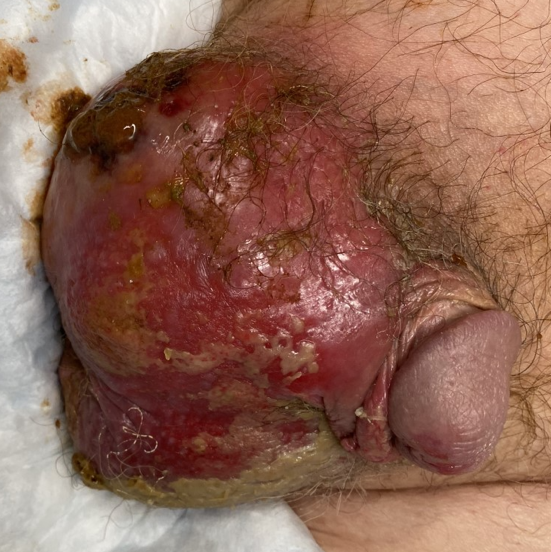
Figure 1: 83-year-old male with right sided ECF from cecum to scrotum. (a) Pre-operative photo taken on admission. (b) Intra-operative photograph with spontaneous scrotal ECF containing bowel. (c) Post-operative photo of right scrotal wound status post debridement, orchiectomy and hernia repair.
The patient was admitted to the ICU, placed on IV antibiotics, and taken to the operating room by general surgery and urology. An incarcerated, right-sided, indirect inguinal hernia containing terminal ileum and the ascending colon was noted, with a 5 cm full-thickness defect in the cecum that fistulized to the right scrotum (Figure 1b). The defect was tied off with silk to prevent further contamination of the abdominal cavity and a formal right hemicolectomy was performed so as to remove any irradiated colon in the pelvis that could potentially compromise an anastomosis. Urology performed a scrotal debridement and right-sided orchiectomy secondary to poor blood supply to the right testicle. General surgery then completed a formal right inguinal herniorrhaphy. The peritoneal defect was closed in two layers without the use of mesh due to fecal contamination.
The scrotum was packed with betadine-soaked gauze, and the patient underwent daily dressing changes (Figure 1c). He endured an uncomplicated post-operative course and returned to his nursing home on post-operative day 10 with home wound care.
Patient 2
The second patient was an 83-year-old male who presented from an assisted living facility after he was noted to have stool pouring from his left scrotum. Past medical history was significant for atrial fibrillation, polio, coronary artery disease, prostate cancer (status post resection many years before; no radiation or chemotherapy), and hypertension. He had been admitted to the hospital two weeks earlier after a fall. At that time, he was noted to have a large left inguinal hernia on CT scan that contained sigmoid in his scrotum. The hernia was easily reducible on physical exam. Since he had no pain or obstructive symptoms, no surgical plans were made at that time.
When he presented to the emergency department two weeks later with stool draining from the left side of his scrotum, his vitals were notable for tachycardia and a leukocytosis of 25.4. On physical exam he was drowsy secondary to septic shock. His abdominal exam revealed peritonitis and a small hole in the left scrotum was seen which had surrounding erythema and was actively draining stool. He was immediately begun on broad spectrum antibiotics and taken emergently to the operating room.
In the operating room, the general surgery team performed an oblique left inguinal hernia incision over the inguinal ligament and immediately noted a large hernia sac. The sac was able to be reduced into the abdomen. The area of sigmoid colon containing the perforation and ECF tract opening was resected. The hernia was repaired using a McVay technique with 2.0 prolene suture. The urology team performed an intra-operative consult and deemed the left hemiscrotum non-viable due to necrosis and gangrene and resected the entire left hemiscrotum. Due to the amount of scrotum removed, the left testicle also had to be resected because of lack of space in the remaining scrotum. General surgery returned to the operating room to create an end colostomy. Using a midline incision, the proximal portion of the stapled off sigmoid was brought out through the LUQ and an end colostomy was created. The left inguinal wound and scrotal wounds were packed with betadine-soaked gauze (Figures 2a and 2b).


Figure 2: 83-year-old male with left sided spontaneous scrotal ECF from sigmoid colon to scrotum. (a) Post-operative photo of scrotal incision where left orchiectomy was performed. (b) Post-operative photo of inguinal incision through which McVay repair performed.
After surgery, the patient was taken to the surgical intensive care unit where he remained intubated and on broad spectrum antibiotics. On post-operative day (pod) 3 he was extubated. His post-operative course was complicated by depression, poor oral intake, and failure to thrive. A percutaneous endoscopic gastrostomy feeding tube was placed on pod 21 and tube feeds reached goal at pod 23. He was discharged to rehab with continued daily dressing changes and wound care on pod 29.
Discussion
After extensive literature review, we believe that our two patients represent the first reported cases of adult spontaneous scrotal ECFs in the United States. The only other case in the literature describing a spontaneous scrotal ECF in the U.S. occurred in an extremely low birth weight infant [5]. The development of a scrotal ECF secondary to an inguinal hernia is extremely rare among adult patients and less than 10 have been identified in the literature worldwide.
The first case of a spontaneous scrotal ECF reported in an adult was in 2006 [6]. Prior to this, all reported cases of spontaneous scrotal ECFs had been in pediatric patients, with the first case reported in 1989 [7]. A case report in 2020, was only the sixth case of a spontaneous ECF involving the scrotum to be published [2,6,8]. Of note, there were three cases involving spontaneous perforation of a Richter’s hernia that led to Fournier’s gangrene in the U.S. in 2007. However, there was no report of a scrotal ECF in any of these three cases [7].
Since the majority of patients in the United States receive prompt medical care for their inguinal hernias, a spontaneous scrotal ECF is quite rare. It is much more commonly seen in countries that have poor access to medical care. The six previously published cases occurred in India, Pakistan, and Nigeria [2,6,8]. Poor access to medical care results in patients presenting later to the hospital. Often, improper medical management occurs due to poverty, neglect, and a lack of medical knowledge [3]. The repair rate of inguinal hernias in the United States is 275 per 100,000 inguinal hernias per year. This is compared to Ghana where the repair rate is estimated to be 30 per 100,000 inguinal hernias. The main contributing factors to the differences in care are the lack of disease education, cost of surgery, cost of anesthesia, and lack of trained surgeons [9].
It is unclear why our two patients took so long to seek medical advice for their scrotal swelling. Of note, the first described patient and his son denied any history of a hernia. However, it does seem unlikely that his ECF developed after only one month of patient described scrotal swelling. What likely occurred in the authors’ opinions was the patient and son did not realize there was a change in his scrotal anatomy, leading to the swelling and hernia going unnoticed. In the second patient, he was aware of having a hernia, especially after his recent hospitalization – however, he likely was unaware of the risks of not having it repaired. Therefore, both patients shared in common not only their age and the fact that they presented from assisted living situations with histories of prostate cancer, but also likely a lack of medical knowledge. The latter was likely the most contributing factor to their delay in hernia treatment and prevention of spontaneous ECF development.
Interestingly, the first patient had an uncomplicated post-operative course unlike the second patient. Although not entirely clear, this is thought to be due to the fact that on initial presentation, the first patient was not septic like the second. The first patient was not peritonitic on his abdominal exam, nor did he have confusion, and he had a significantly lower leukocytosis. However, the first patient did have subcutaneous emphysema in his scrotum. It appears that the first patient had a somewhat controlled perforation to his scrotum versus the second patient who was peritonitic on exam secondary to uncontained perforation into his abdomen.
As previously mentioned, the first patient did have a history of brachytherapy for bladder cancer several years prior, as well as radiation for prostate cancer over five years before. The literature cites that 75% of patients undergoing abdominal or pelvic radiation will suffer some type of radiation enteritis, which commonly presents as a stricture or bleeding. In addition, a small subset of patients will develop fistulous complications that usually take about six months to become apparent. Furthermore, cytoreductive surgery and intraperitoneal hyperthermic chemotherapy have been linked in some cases to the development of multiple ECFs. In our research, we were unable to find any links in the literature between brachytherapy and ECFs [10]. In addition, this patient was also on daily prednisone and during our literature review we did not note any link between steroid use and ECF development. However, his daily prednisone use did likely play a role in his overall septic presentation.
We conclude that our first patient’s scrotal ECF was spontaneous in nature and solely a result of his incarcerated inguinal hernia. The second patient had no known risk factors for development of an ECF and as noted he was hospitalized two weeks prior and the hernia was reducible. This leads us to the conclusion that he also developed a spontaneous scrotal ECF.
Conclusion
To our knowledge, these are the first recorded cases describing an adult spontaneous scrotal ECF in the United States and the seventh and eighth reported cases worldwide. It is unclear why the first described patient took so long to seek medical advice, although he denied any history of hernia and only noted scrotal swelling for one month prior to presentation. The other patient was deemed to have an asymptomatic, reducible hernia two weeks prior and was not in need of emergent repair. Both patients likely had delayed presentations of their incarcerated hernias as their scrotal ECFs decompressed the bowels and attenuated the development of obstructive symptoms. Both patients ultimately underwent a successful orchiectomy by urology, in addition to bowel resection with ligation of their scrotal ECFs, and herniorrhaphy by general surgery.
References
2. Asghar MS, Kaleem A, Rijal S. A rare case of spontaneous scrotal enterocutaneous fistula as a complication of incarcerated inguinal hernia with review of literature. The Professional Medical Journal. 2020 Jul 10;27(07):1537-40.
3. Koshariya M, Naik S, Rai A. Incarcerated inguinal hernia presenting as spontaneous scrotal fecal fistula. Hernia. 2006 Oct;10(5):434-5.
4. Malik P, Rathi M, Kumar K, Sharma R, Meena P, Arya A, et al. Scrotal enterocutaneous fistula: a rare initial presentation of inguinal hernia. Journal of Surgical Case Reports. 2014 Jun 1;2014(6).
5. Smith CA, McAdams RM, Lisse G, Avansino JR. Scrotal enteric fistula in an extremely low birth weight premature neonate. Journal of Pediatric Surgery Case Reports. 2018 Mar 1;30:44-5.
6. de Campos-Lobato LF, Vogel JD. Enterocutaneous fistula associated with malignancy and prior radiation therapy. Clinics in colon and rectal surgery. 2010 Sep;23(03):176-81.
7. Orangio GR. Enterocutaneous fistula: medical and surgical management including patients with Crohn's disease. Clinics in colon and rectal surgery. 2010 Sep;23(03):169-75.
8. Bahadur A, Singh N, Kashmira M, Shukla A, Gupta V, Jain S. Fecal scrotal abscess secondary to spontaneous retroperitoneal perforation of ascending colon. Case Reports in Medicine. 2021 Mar 29;2021.
9. Amoako JK, Wordui T, Kumassah PK, Amissah JM, Ohene-Yeboah M. Entero-scrotal fistula in an adult: a complication of traditional health care practice in Ghana. Clinics in Surgery. 2018;3(1):2025.
10. Ahi KS, Moudgil A, Aggarwal K, Sharma C, Singh K. A rare case of spontaneous inguinal faecal fistula as a complication of incarcerated Richter’s hernia with brief review of literature. BMC surgery. 2015 Dec;15(1):1-5.