Commentary
Periungual capillaroscopy is a technique that allows the study of the microcirculation of the periungual beds in a non-invasive and simple way, allowing the papillary dermis to be visualized in vivo [1]. It is one of the fundamental diagnostic tests in clinical practice and teaching in rheumatology today.
The basic patterns in capillaroscopy are determined by the structure and architecture of the nail bed, capillary density, the presence of avascular areas, capillary morphology and vascular flow [1]. Normal capillaroscopy pattern is mainly characterized by a hairpin arrangement of capillaries with a regular "comb-like" distribution, the presence of one capillary in each dermal papilla and the homogeneity in the morphology of the capillaries observed in the microscopic field (Figure 1).
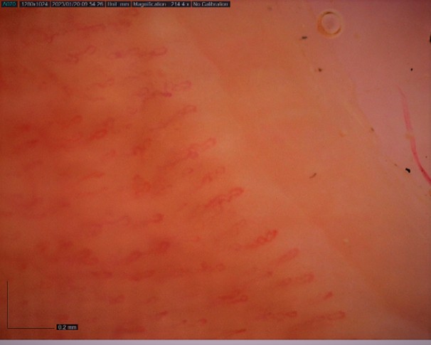
Figure 1. Capillaroscopic image corresponding to normality pattern.
Currently the main indication for capillaroscopy is initial assessment of patients with Raynaud's phenomenon, which is present in many autoimmune rheumatic diseases, especially scleroderma. Firstly, it allows differentiation between patients who present the phenomenon in isolation (primary Raynaud's) and those in whom it appears in the context of an autoimmune disease (secondary Raynaud's) [2]. In the latter, the phenomenon usually appears very early in the development of these diseases, and may even precede the onset of these diseases by years, making early diagnosis essential [3].
The capillaroscopic pattern suggestive of scleroderma is characterized by the observation of megacapillaries, hemorrhages in drops or bands, disorganization of the capillary bed, avascular areas and newformed capillaries (Figure 2).
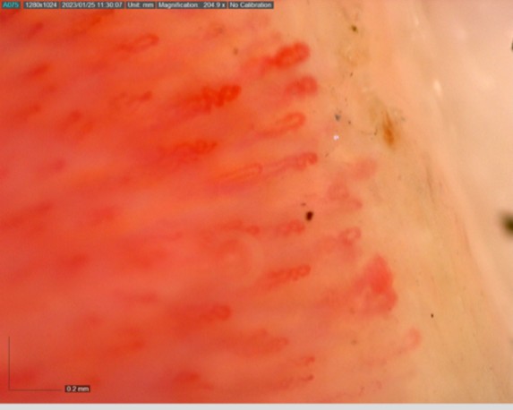
Figure 2. Capillaroscopic image showing characteristic changes of scleroderma, such as megacapillaries and microhemorrhages.
From a technical point of view, nail bed capillaroscopy can be performed with different types of optical devices, such as the handheld dermatoscope, the digital epiluminescence microscope or the optical microscope with adapted camera. The most widely used in the clinic was the so-called stereomicroscope, which is an optical binocular microscope adapted to a camera [1].
In recent years, a new technique called nailfold videocapillaroscopy (NVC) [6] has been developed which has a number of practical advantages over previous techniques. The video-capillaroscope is the result of incorporating hardware and software suitable for digitizing images into the optical microscope normally used. It is based on the attachment of a video camera to the microscope to obtain high-resolution images of the microcirculation of the nail bed (752 x 582 pixels) at a magnification up to 1000x which allows a more detailed observation of the periungueal bed, facilitating the correct diagnosis. Images at magnifications close to 1000x even allow observation of the passage of erythrocytes through the capillaries.
One of the main differences between steromicrocopy and NVC is that the latter allows the images to be stored on a computer for further processing using different software programs (Table 1). Among the most used are the following:
| AUTOCAPI | Automatic count of total number of capillaries |
| MANCHESTER SISTEM | Possibility of obtaining mosaic images |
| CAPILLARY. IO | Website that allows to analyze any capillaroscopy image with artificial intelligence |
| VISION TRANSFORMER | Classification of image into a specific microangiopathy pattern |
The so-called "AUTOCAPI" [6,7], developed by Cutolo et al. in 2017, is a software that automatically counts the total number of capillaries in the nail bed, constructed, through an exploratory image set. The authors created application rules to define the region of interest in the images.
The "Manchester Sistem" [8,9], which is a software-controlled video microscope with a motorized mount, developed in 2019 by Berks et al. This software, in addition to counting the number of capillaries, offers advantages over previous applications, such as the possibility of obtaining mosaic images, and, above all, the automatic measurement of capillary flow.
The Capillary.io application [10,11] has been developed in 2021 by Gracia-Tello et al. This system provides rheumatologists with a web page where they can enter their capillaroscopic images, which are processed automatically through artificial intelligence, and can analyze any capillaroscopy image, regardless of the optical device used to obtain it. It allows automatic measurement of capillary size, and interprets the whole image, allowing the detection of qualitative alterations, such as the presence of hemorrhages or morphologically altered capillaries (Figure 3).
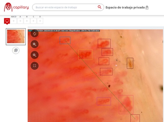
Figure 3. Computer-assisted capillaroscopic imaging using the Capillary.io application.
The Vision Transformer (ViT) is a software [12,13] developed in 2022 by Garaiman et al. It is an artificial intelligence application that automatically classify any capillaroscopy image into a specific microangiopathy pattern, such as the sclerodermal pattern. This automatic classification is the major contribution of this innovation to the field. Method is based in some parameter analysis, such as capillary diameter and size, avascular areas and the presence or absence of microhemorrhages.
The different software programs allows the automatic performance of different measurements on the images obtained that are essential for diagnosis, such as dimensions and diameters of the capillary structures, accounting for the number of capillaries (capillary density), hemorrhages detection in the scanned area, automatic classification of capillaries into normal, tortuous, or branched capillaries, presence of signs of neovascularization on the images and blood flow through the blood vessels in the studied area, even being able to identify blood elements inside them. In this sense, videocapillaroscopy allows the visualization of images in movement, providing, unlike previous techniques, a dynamic and functional study.
The automatic measurement process facilitates and speeds up the rheumatologist's work, as the measurements do not have to be performed manually, which optimizes patient classification, favoring comparative, longitudinal and multicenter studies [10].
Another great advantage of NVC is that it is an easy technique that can be performed quickly in the rheumatologist's own office, even in the first assessment of a patient with Raynaud's phenomenon, which speeds up the diagnostic process while reducing the possibility of ordering unnecessary repeated tests in patients who probably would not develop autoimmune diseases.
NVC offers the possibility of automatic storage of the images obtained in the computer, facilitating their subsequent computer processing, which, in addition to facilitating further visualization for a correct diagnosis, it offers advantages for monitoring the evolution of patients over time. In the field of teaching, the availability of images in computer files facilitates the development of teaching materials, such as slide presentations, videos or learning applications. The evolutionary explanation of these images is key to the understanding, by doctors in training, of the appropriate diagnostic classification of rheumatological patients.
The digitalization of capillaroscopy and the continuous improvement of its optical systems has allowed, in recent years, to expand its indications and usefulness beyond the initial evaluation of patients with Raynaud's phenomenon.
In this sense, NVC optimizes the patient follow-up process and allows monitoring of disease progression and detection of significant changes in patients' capillaroscopic patterns. An example of this is the observational study by Avouac et al. [11] which concludes that sequential performance of this technique is suitable for monitoring scleroderma, noting that progressive capillary loss over time is a marker of disease progression. NVC may have predictive value of new disease manifestations and complications. Thus, the 2017 study by Markusse et al. [12] concluded that the more severe the pattern observed with NVC, the higher is the likelihood of developing severe organ involvement in patients with scleroderma. The NVC patterns were significantly associated with severe pulmonary and vascular involvement several months after the scan. In addition, the pattern observed with NVC showed a stable association with the presence of interstitial lung disease or pulmonary hypertension. More recently, a 2022 study by Umashankar et al. concluded that NVC can increase the earlier diagnosis of interstitial lung disease in patients with scleroderma and can help determine its classification [13].
In recent years, NVC has begun to be used in rheumatological diseases other than scleroderma, such as Behçet's disease and vasculitis. Regarding Behçet's syndrome, in the study of Mercadé Torras et al., it is concluded that non-specific abnormalities of microcirculation are common in patients with this syndrome, and presence of ≥2 non-specific qualitative abnormalities in NVC is associated with vascular manifestations, especially with thrombotic events [14]. Regarding vasculitis, the study by Matsuda et al. concludes that in patients with ANCA-associated vasculitis NVC abnormalities are significantly associated with disease severity, which suggests that NVC is a useful tool for assessing the disease activity and treatment response [15].
NVC is also beginning to be used in the assessment of childhood rheumatic conditions. In the 2023 study by Melsens et al., a significant difference was found between the NVC patterns of children with rheumatic diseases and those of healthy controls. A lower capillary density and a higher number of dilated and giant capillaries were observed in children with juvenile systemic sclerosis, mixed connective tissue disease, and juvenile dermatomyositis than in children without rheumatic pathology. The authors conclude that NVC should be performed regularly in children with primary Raynaud's phenomenon and localized scleroderma [16].
Finally, research is currently being conducted into the possible application of NVC in non-rheumatological pathologies, such as cardiovascular diseases. NVC is an established, noninvasive, easily applicable technique for the assessment of peripheral microcirculation. Therefore, capillaroscopic findings in patients with cardiovascular risk factors could be correlated with clinical and laboratory markers of cardiac function [17].
In conclusion, the NVC accelerates the diagnostic process of scleroderma, optimizes patient monitoring and monitors the progression of the disease, allowing to detect complications such as pulmonary affectation in initial phases. The application of artificial intelligence can allow in the future to further improve the benefits of this technique.
More research is necessary to determine the usefulness of NVC in other rheumatological diseases such as Behçet's syndrome and vasculitis, as well as its application in pediatric rheumatology and in non-rheumatological pathologies.
References
2. Lambova SN, Müller-Ladner U. The role of capillaroscopy in differentiation of primary and secondary Raynaud's phenomenon in rheumatic diseases: a review of the literature and two case reports. Rheumatol Int. 2009 Sep;29(11):1263-71.
3. Alperi López M. Manual SER de enfermedades reumáticas ((6ª edición). Barcelona, España: Editorial Elsevier; 2014.
4. Movasat A, Shahram F, Carreira PE, Nadji A, Akhlaghi M, Naderi N, et al. Nailfold capillaroscopy in Behçet's disease, analysis of 128 patients. Clin Rheumatol. 2009 May;28(5):603-5.
5. Corominas H, Ortiz-Santamaría V, Castellví I, Moreno M, Morlà R, Clavaguera T, et al CapiCAT group. Nailfold capillaroscopic findings in primary Sjögren's syndrome with and without Raynaud's phenomenon and/or positive anti-SSA/Ro and anti-SSB/La antibodies. Rheumatol Int. 2016 Mar;36(3):365-9.
6. Cutolo M, Trombetta AC, Melsens K, Pizzorni C, Sulli A, Ruaro B, et al. Automated assessment of absolute nailfold capillary number on videocapillaroscopic images: Proof of principle and validation in systemic sclerosis. Microcirculation. 2018 May;25(4):e12447.
7. Berks M, Tresadern P, Dinsdale G, Murray A, Moore T, Herrick A, et al. An automated system for detecting and measuring nailfold capillaries. InMedical Image Computing and Computer-Assisted Intervention–MICCAI 2014: 17th International Conference, Boston, MA, USA, September 14-18, 2014, Proceedings, Part I 17 2014 (pp. 658-665). Springer International Publishing.
8. Gracia Tello B, Ramos Ibañez E, Fanlo Mateo P, Sáez Cómet L, Martínez Robles E, Ríos Blanco JJ, et al. The challenge of comprehensive nailfold videocapillaroscopy practice: a further contribution. Clin Exp Rheumatol. 2022 Oct;40(10):1926-32.
9. Garaiman A, Nooralahzadeh F, Mihai C, Gonzalez NP, Gkikopoulos N, Becker MO, Distler O, Krauthammer M, Maurer B. Vision transformer assisting rheumatologists in screening for capillaroscopy changes in systemic sclerosis: an artificial intelligence model. Rheumatology (Oxford). 2023 Jul 5;62(7):2492-2500.
10. Gronenschild EHBM, Muris DMJ, Schram MT, Karaca Ü, Stehouwer CDA, Houben AJHM. Semi-automatic assessment of skin capillary density: Proof of principle and validation. Microvasc Res. noviembre de 2013;90:192-8.
11. Avouac J, Lepri G, Smith V, Toniolo E, Hurabielle C, Vallet A, et al. Sequential nailfold videocapillaroscopy examinations have responsiveness to detect organ progression in systemic sclerosis. Semin Arthritis Rheum. 2017 Aug;47(1):86-94.
12. Markusse IM, Meijs J, de Boer B, Bakker JA, Schippers HPC, Schouffoer AA, et al. Predicting cardiopulmonary involvement in patients with systemic sclerosis: complementary value of nailfold videocapillaroscopy patterns and disease-specific autoantibodies. Rheumatology (Oxford). 2017 Jul 1;56(7):1081-8.
13. Umashankar E, Abdel-Shaheed C, Plit M, Girgis L. Assessing the role for nailfold videocapillaroscopy in interstitial lung disease classfication: a systematic review and meta-analysis. Rheumatology (Oxford). 2022 May 30;61(6):2221-34.
14. Mercadé-Torras JM, Guillén-Del-Castillo A, Buján S, Solans-Laque R. Nailfold videocapillaroscopy abnormalities and vascular manifestations in Behçet's syndrome. Clin Exp Rheumatol. 2024 Oct;42(10):2065-70.
15. Matsuda S, Kotani T, Wakura R, Suzuka T, Kuwabara H, Kiboshi T, et al. Examination of nailfold videocapillaroscopy findings in ANCA-associated vasculitis. Rheumatology (Oxford). 2023 Feb 1;62(2):747-57.
16. Melsens K, Cutolo M, Schonenberg-Meinema D, Foeldvari I, Leone MC, Mostmans Y, et al. EULAR Study Group on Microcirculation in Rheumatic Diseases. Standardized nailfold capillaroscopy in children with rheumatic diseases: a worldwide study. Rheumatology (Oxford). 2023 Apr 3;62(4):1605-15.
17. Baroutidou A, Arvanitaki A, Pagkopoulou E, Anyfanti P, Ziakas A, Kamperidis V, et al. Nailfold videocapillaroscopy as a non-invasive tool for the assessment of peripheral microangiopathy in cardiovascular diseases. J Hypertens. 2025 Jan 1;43(1):48-65.