

Abstract
Pleckstrin Homology-like Domain family B member 3 (PHLDB3) is a member of the PHLDB protein family characterized by the presence of two central coiled-coil (CC) domains and a C-terminal pleckstrin homology (PH) domain. This family includes PHLDB1 with 1377 amino acids (AAs), PHLDB2 with 1253 AAs, and PHLDB3 with 640 AAs. Although more studies have been reported on PHLDB1 and PHLDB2, our lab has made some progress in investigating PHLDB3. We previously found that PHLDB3 participates in the MDM2-p53 negative feedback loop. Recently, our study showed that PHLDB3 also plays a regulatory role in mediating the mTORC2-AKT1 signaling. Hence, this minireview will focus on discussing this recent discovery of PHLDB3, its potential implications and biological significance in addition to briefly reconciling this pathway with its oncogenic role via p53 inactivation.
Keywords
PHLDB3, mTORC2, AKT1, Liprin-α1, Signal transduction, Colorectal cancer, Metabolism
Introduction
Among the three PHLDB family members (PHLDB1, PHLDB2 and PHLDB3), PHLDB3 has been the least studied [1]. Over the past several years, our lab has made significant progress toward understanding its biological and biochemical functions. Previously, our lab identified PHLDB3 as a transcriptional target of p53 and showed that it assists MDM2-mediated p53 degradation, thereby attenuating p53 tumor suppressor activity and promoting tumor growth [2]. Subsequent studies by us and others demonstrated that PHLDB3 contributes to tumor progression in several cancer types [2,3], suggesting that its biological functions extend beyond the canonical p53 pathway. Indeed, our latest study revealed that PHLDB3 can bind to RICTOR, a subunit of the mechanistic target of rapamycin complex 2 (mTORC2), thereby enhancing mTORC2-AKT signaling and promoting colorectal cancer growth [4]. mTORC2 is a highly conserved protein kinase complex composed of mTOR, RICTOR, mSIN1, and mLST8 [5]. As a central regulator of AKT Ser473 phosphorylation, mTORC2 plays critical roles in cell survival, proliferation, metabolism, and cytoskeletal organization [6,7]. Dysregulation of mTORC2 signaling has been implicated in a wide range of human diseases, particularly cancer and metabolic disorders [8].
Although PHLDB3 and mTORC2 were originally studied in distinct signaling contexts, our recent findings linked them by identifying RICTOR as a novel PHLDB3-interacting protein and demonstrating that PHLDB3 promotes mTORC2-dependent AKT activation [4]. These observations expand the functional landscape of PHLDB3 and position it as a previously unrecognized regulator of mTORC2 signaling. Because the biological functions and disease associations of PHLDB1 and PHLDB2 have been discussed in detail in our previous review [1], the present mini-review, in addition to briefing the current understanding of PHLDB3 biology, primarily focuses on discussion of our recent advances linking PHLDB3 to mTORC2-AKT signaling as well as broader implications of this signaling axis in cancer progression and metabolic regulation. We also envision the potential therapeutic opportunities arising from the emerging role of PHLDB3 in mTORC2 regulation.
The mTORC2 Signaling Pathway
Among the four subunits of the mTORC2 kinase complex as mentioned above [5], RICTOR serves as a defining scaffold protein that distinguishes mTORC2 from mTORC1 and is required for complex assembly and substrate recognition [9,10]. mSIN1 contains a pleckstrin homology domain that contributes to membrane localization and growth factor responsiveness of the complex [11]. Upon growth factor stimulation, phosphatidylinositol 3-kinase (PI3K) signaling promotes the recruitment and activation of mTORC2 at cellular membranes [12]. Activated mTORC2 phosphorylates several AGC family kinases, including AKT, SGK, and PKC [13]. Among these substrates, phosphorylation of AKT at Ser473 represents one of the best-characterized functions of mTORC2 and is required for maximal AKT activation [6]. Through AKT and other downstream effectors, mTORC2 regulates cell survival, proliferation, glucose metabolism, lipid metabolism, and cytoskeletal dynamics [14].
Although the downstream functions of mTORC2 have been extensively investigated, the mechanisms underlying its activation and regulation remain incompletely understood. Previous studies have identified growth factor signaling, phosphoinositides, and membrane localization as important regulatory factors [12]. However, a complete picture of the mTORC2 pathway remains to be fully unveiled. Identifying yet unknown regulators of the RICTOR-mTORC2 complex is therefore important for better understanding how mTORC2 signaling is controlled in normal physiology and pathological alterations including cancer.
Our recent study has begun to uncover previously unrecognized mechanisms regulating mTORC2 activity. Specifically, we found that PHLDB3 directly interacts with RICTOR, consequently promoting mTORC2-dependent AKT activation [4]. This finding raises the possibility that PHLDB3 functions as an upstream regulator of mTORC2 signaling and provides a new direction for investigating the biological functions of the understudied protein PHLDB3.
PHLDB3
Although PHLDB1, PHLDB2, and PHLDB3 share the characteristic PH domain, which is a structural module often associated with phosphoinositide binding and membrane-related signaling events [15], accumulating evidence suggests that individual family members may participate in distinct cellular processes. PHLDB1 and PHLDB2 have been implicated in several biological and disease-related contexts, as summarized in detail in our previous review [1], whereas the mechanistic studies of PHLDB3 have primarily linked it to the MDM2–p53 feedback loop and, more recently, mTORC2–AKT signaling [2,4]. As mentioned above, compared with other PHLDB family members, PHLDB3’s biochemical activity and biological functions remain largely obscure. Initially, our lab discovered the PHLDB3-encoding gene as a p53-inducible gene while screening for p53 transcriptional targets [2]. Briefly, upon p53 activation, PHLDB3 is transcriptionally induced and subsequently enhances MDM2-mediated ubiquitination and degradation of p53. Through this negative feedback mechanism, PHLDB3 attenuates p53 tumor suppressor activity and supports cancer cell growth [2]. These findings establish PHLDB3 as an oncoprotein by suppressing the tumor suppressor function of p53 (Figure 1). During this study, we observed that PHLDB3 may also participate in p53-independent oncogenic processes, as depletion of PHLDB3 suppressed proliferation of p53-null colorectal cancer cells as well. As discussed further below, this observation led to our recent discovery of PHLDB3 as a player in regulation of mTORC2-AKT signaling independently of p53 [2,4].
The Liprin-α1-PHLDB3-mTORC2-AKT Signaling
To identify other potential signaling partners of PHLDB3, we first performed proteomic analyses of PHLDB3-associated proteins in HEK293T cells expressing FLAG-PHLDB3 using co-immunoprecipitation (co-IP) coupled with liquid chromatography–tandem mass spectrometry [4]. Among the proteins identified, RICTOR topped out as a prominent candidate [4]. Our subsequent co-immunoprecipitation and protein-protein interaction assays confirmed the interaction between PHLDB3 and RICTOR in cells and in vitro. Domain-mapping studies demonstrated that the 1st coiled coil domain (CC1) of PHLDB3 is essential for RICTOR binding. These studies established a physical association between PHLDB3 and RICTOR, a subunit of the mTORC2 complex.
Functional analyses further demonstrated that PHLDB3 acts as a positive regulator of mTORC2 activity [4]. Silencing PHLDB3 markedly reduced AKT phosphorylation at Ser473, whereas ectopic expression of PHLDB3 enhanced mTORC2-dependent AKT activation [4]. Given that AKT Ser473 phosphorylation represents a hallmark of mTORC2 signaling [6], these findings support the notion that PHLDB3 functions as a positive regulator of the mTORC2-AKT pathway.
Remarkably, our further studies revealed Liprin-α1 as another upstream regulator of the PHLDB3-mTORC2-AKT pathway [4]. First, Liprin-α1 associates with PHLDB3 in the cytoplasm of cells, and this association was confirmed by co-immunoprecipitation [4]. Also, Liprin-α1 acts to stabilize the PHLDB3 protein, as depletion of Liprin-α1 led to the decreased PHLDB3 abundance and half-life, attenuated AKT phosphorylation, and suppressed tumor cell growth [4]. Consistently, ectopic expression of Liprin-α1 increased PHLDB3 protein levels by blocking proteasome-mediated degradation, thereby enhancing AKT phosphorylation [4], although the E3 ubiquitin ligase responsible for PHLDB3 degradation remains to be identified. These observations support a hierarchical Liprin-α1–PHLDB3–RICTOR–mTORC2 signaling axis and reveal an additional regulatory layer governing mTORC2 activity (Figure 1).
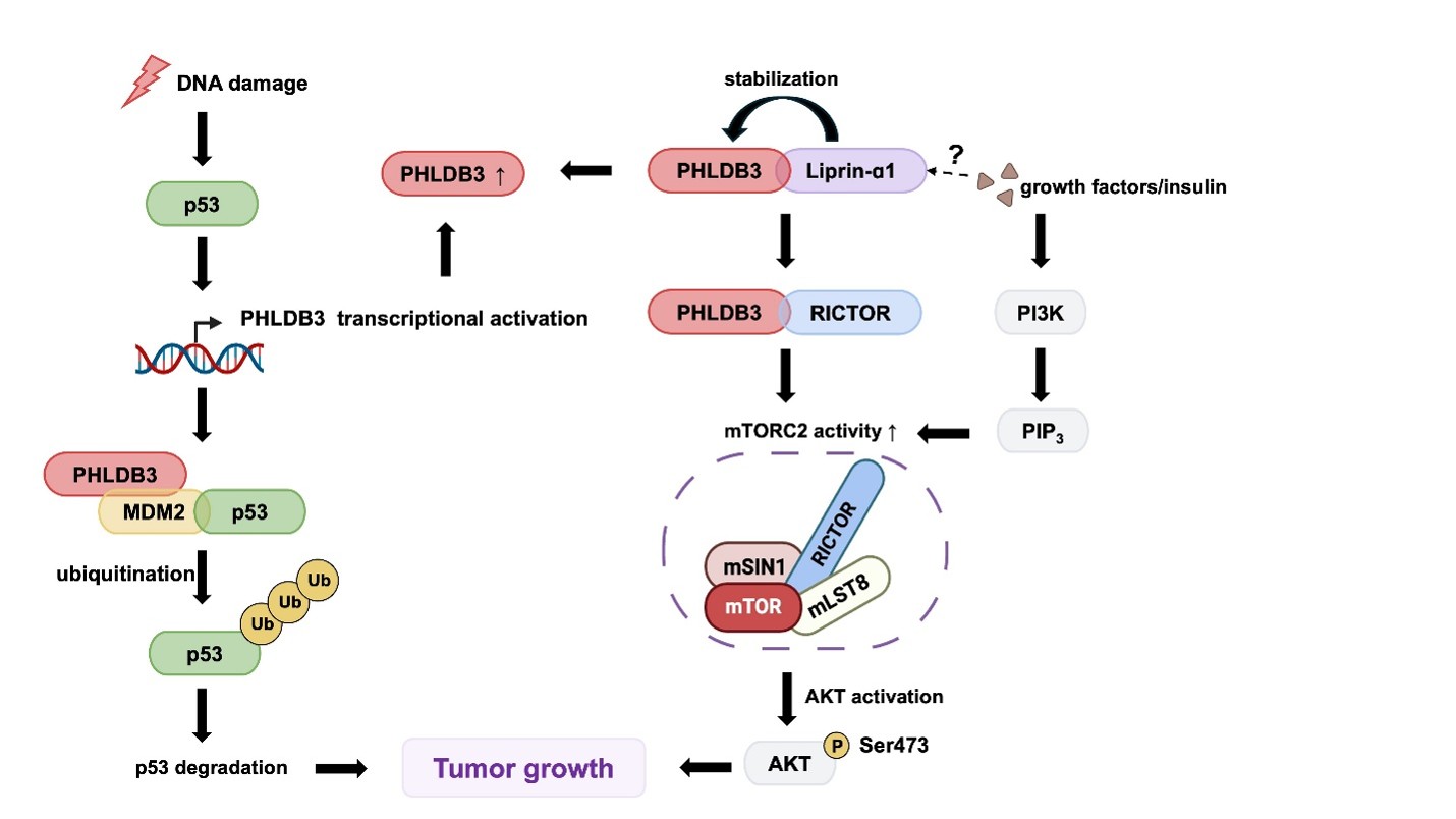
Collectively, these studies redefine PHLDB3 as more than a downstream effector of p53. Instead, PHLDB3 can also act as an upstream regulator of mTORC2 that integrates multiple signaling pathways through its interaction with RICTOR, though the precise molecular mechanism by which PHLDB3 regulates the activity of the mTORC2 kinase still needs to be further dissected. The discovery of the PHLDB3–RICTOR–mTORC2 axis not only broadens our understanding of PHLDB3 biology but also provides a conceptual framework for investigating its roles in cancer, metabolism, and other physiological and pathological processes. Of note, although the evidence defining PHLDB3 as a regulator of the RICTOR-mTORC2-AKT pathway has thus far been reported only from our recent study [4], the results described in the report have been independently reproduced by multiple researchers in our lab. Additional studies using different cellular, organoid, and animal model systems will be necessary and important to further consolidate the concept of this signal pathway and its biologicant significance.
Biological Implications of the Liprin-α1-PHLDB3-mTORC2-Akt Axis
Through its ability to regulate mTORC2-dependent AKT activation, the Liprin-α1–PHLDB3–mTORC2 signaling axis may influence a broad spectrum of biological and pathological processes.
Our findings established the oncogenic role of this axis in colorectal cancer (CRC). Several lines of evidence support this assumption. First, PHLDB3 is frequently upregulated in human colorectal tumors and associated with advanced disease and poor clinical outcomes [2,4]. Also, it appears to promote CRC cell growth via two distinct pathways: Chao et al. identified PHLDB3 as a negative regulator of the tumor suppressor function of p53 through its interaction with MDM2 [2], whereas Ko et al. demonstrated that PHLDB3 also promotes oncogenic mTORC2 signaling in colorectal cancer cells [4]. In Liprin-α1-depleted CRC models, PHLDB3 re-expression restored AKT phosphorylation and rescued growth phenotypes [4]. The identification of Liprin-α1 as an upstream regulator further extends this signaling network. By stabilizing PHLDB3 and facilitating mTORC2 activation, Liprin-α1 provides an additional regulatory layer that supports sustained oncogenic signaling. Consistently, Liprin-α1 is highly expressed in CRCs and associated with poor survival of CRC patients [4]. Collectively, these findings support the Liprin-α1–PHLDB3–mTORC2 axis as a critical signaling module driving CRC progression, complementing the previously established role of PHLDB3 in suppressing p53 activity (Figure 1) and suggest that multiple components of this pathway may represent potential therapeutic targets. It would be interesting to investigate whether PHLDB3 plays an oncogenic role in other types of cancers, such as lung, breast, and liver cancers.
In addition to its oncogenic role, preliminary observations have raised the possibility that PHLDB3 may also play biological roles in other aspects. A quantitative proteomic analysis of human dental pulp stem cells identified PHLDB3 as one of the most highly upregulated proteins during odontogenic differentiation, indicating a potential role in tissue development and regeneration [16]. However, a causal role for PHLDB3 in this process has not been experimentally tested. This represents another future direction for PHLDB3 research. Furthermore, integrative genomic analyses have identified PHLDB3 as a candidate gene shared between Alzheimer's disease and sarcopenia, with predicted expression changes in both skeletal muscle and brain tissues [17], well consistent with its positive role in the mTORC2-AKT signaling. Although these associations are bioinformatic and correlative and should not be interpreted as evidence that PHLDB3 functionally contributes to either disorder, they imply that PHLDB3 may participate in diverse physiological processes outside of cancer. Further experimental studies using appropriate cellular and animal models will be conducive to determining whether these observations have biological significance.
A potential, yet unvalidated, implication of the PHLDB3–mTORC2 connection concerns metabolic homeostasis. In addition to promoting cell survival and proliferation, mTORC2 serves as a central regulator of glucose uptake, insulin signaling, lipid metabolism, and cellular energy balance through activation of AKT, SGK1, and other AGC family kinases [18]. Consequently, dysregulated mTORC2 signaling has been implicated in obesity, insulin resistance, type 2 diabetes, non-alcoholic fatty liver disease, and other metabolic disorders [19]. Because PHLDB3 functions upstream of mTORC2, its biological influence may extend beyond tumorigenesis to physiological processes governed by mTORC2. Although direct evidence linking PHLDB3 to metabolic disease is currently lacking, regulation of mTORC2 by PHLDB3 raises the possibility that PHLDB3 contributes to insulin responsiveness, glucose utilization, and metabolic adaptation under specific physiological or pathological conditions. This conjecture should be regarded as an open question that will require direct testing, particularly in metabolically relevant tissues and animal models .
Interestingly, PHLDB3 contains a pleckstrin homology (PH) domain, a structural feature commonly involved in phosphoinositide-dependent membrane signaling [1]. Since phosphoinositides are central mediators of insulin signaling and have emerged as important contributors to mTORC2 activation [11,20], one testable hypothesis is that the PH domain of PHLDB3 participates in phosphoinositide-dependent localization or signaling. Although this hypothesis remains to be experimentally tested, it provides a potential mechanistic framework for understanding how PHLDB3 may coordinate growth factor signaling, cellular metabolism, and mTORC2 activation.
Therapeutic Implications of Targeting the Liprin-α1-PHLDB3-mTORC2 Signaling Axis
Originally recognized as an MDM2-binding protein, PHLDB3 has recently been identified as a direct binding partner of RICTOR, substantially expanding our understanding of its biological functions. Rather than functioning as an isolated signaling protein, PHLDB3 may serve as a signaling scaffold by coordinating multiple protein interactions (Figure 1). This unique positioning places PHLDB3 at a critical regulatory node where p53-mediated stress responses converge with PI3K–AKT–mTOR signaling. This creates opportunities for therapeutic intervention beyond conventional kinase inhibition.
Unlike catalytic kinases, scaffold proteins regulate signaling by controlling protein complex assembly, spatial organization, and pathway specificity [21]. These properties make scaffold-dependent signaling conceptually attractive for therapeutic intervention, but scaffold proteins also present substantial drug-development challenges. In particular, protein-protein interaction surfaces are often large, relatively flat, and dynamic and may lack well-defined small-molecule-binding pockets, complicating the development of potent and selective inhibitors [22]. In addition, because scaffold proteins can engage multiple partners in a context-dependent manner, disrupting one interaction may produce incomplete or unanticipated pathway effects [23]. Thus, PHLDB3 should currently be viewed as a candidate therapeutic target, whose druggability and therapeutic window remain to be determined.
Several therapeutic approaches could be envisioned for targeting this newly identified signaling axis. The first strategy is selective inhibition of mTORC2 signaling. Aberrant activation of mTOR signaling has long been recognized as an attractive therapeutic target in cancer [24]. However, most clinically available mTOR inhibitors primarily target mTORC1, whereas selective inhibition of mTORC2 remains challenging because both complexes share the same catalytic kinase [25]. Since PHLDB3 specifically promotes RICTOR-dependent mTORC2 activation, targeting PHLDB3 may provide an alternative means of attenuating mTORC2 activity without completely blocking mTOR kinase function. A second strategy involves disrupting the interaction between PHLDB3 and RICTOR. Increasing evidence suggests that modulation of protein–protein interactions represents a feasible therapeutic approach for signaling scaffold proteins. Structural and biochemical characterization of the PHLDB3–RICTOR interface will first be required to determine whether the interaction contains a tractable binding hotspot. Even if such a site is identified, achieving sufficient affinity, selectivity, intracellular delivery, and pharmacological stability may be challenging, particularly for peptide-based inhibitors [22]. Finally, direct elimination of PHLDB3 or Liprin-α1 could represent an alternative strategy, although this idea also requires substantial experimental validation. Because PHLDB3 functions upstream of mTORC2 while simultaneously participating in p53 regulation, inhibition of PHLDB3 may suppress oncogenic AKT signaling while restoring p53 tumor suppressor activity in tumors retaining wild-type TP53. Targeting Liprin-α1 could also post a one-stone with two birds effect, because Liprin-α1 has been shown to play a key role in cancer metastasis [26-28]. Although no specific PHLDB3 or Liprin-α1 inhibitors have yet been reported or in clinical trials, emerging targeted protein degradation technologies, including proteolysis-targeting chimeras (PROTACs), molecular glue degraders, and PHLDB3- or Liprin-α1-specific microRNAs or siRNAs, may provide feasible approaches for selectively removing PHLDB3 or Liprin-α1 protein or decreasing its expression in the future, but their feasibility, specificity, delivery, and safety in vivo remain to be established.
In addition to its therapeutic potential, PHLDB3 may also serve as a clinically useful biomarker. Elevated PHLDB3 expression has been associated with enhanced mTORC2-AKT signaling and colorectal cancer progression, suggesting that PHLDB3 expression levels may reflect pathway activation and identify patients who are more likely to benefit from therapies targeting the PI3K–AKT–mTOR pathway.
Beyond CRC, bioinformatic analyses of breast cancer cohorts demonstrated that PHLDB3 expression correlates with molecular subtype, TP53 status, patient survival, and response to endocrine and anti-HER2 therapies [3]. A recent integrative transcriptomic study also identified PHLDB3 as one of five genes comprising an oxaliplatin resistance-associated prognostic signature in colorectal cancer. Patients with higher-risk signatures exhibited significantly worse survival, and PHLDB3 expression was experimentally confirmed to change following oxaliplatin treatment in multiple CRC cell lines, supporting its potential involvement in chemotherapy response [29]. Together, these findings indicate that PHLDB3 has the potential to serve not only as a prognostic biomarker but also as a predictive biomarker for therapeutic response. Nevertheless, prospective clinical studies involving larger patient cohorts will be required to validate its clinical applications.
Questions and Perspectives
Despite recent advances, much more needs to be learned about the biochemical functions and biological roles of PHLDB3 in cancer development and physiology. Specifically, several important areas warrant further investigation beyond those discussed above. First, the structural basis underlying the interaction between PHLDB3 and RICTOR remains unknown. High-resolution structural studies using cryo-electron microscopy (EM) or X-ray crystallography will be essential for defining the interaction interface and enabling structure-guided development of inhibitors targeting this signaling complex. Such biophysical studies would allow us to further elucidate the atomic and molecular mechanisms underlying the PHLDB3 regulation of mTORC2 activity. The knowledge gained from these studies would further facilitate our future drug discovery targeting the PHLDB3-RICTOR interaction. Second, although PHLDB3 contains a PH domain, the physiological function of this domain still needs to be clarified. Given that PH domains commonly mediate phosphoinositide-dependent membrane recruitment, it will be important to determine whether PHLDB3 responds to phosphoinositide signaling and whether this mechanism contributes to mTORC2 activation.
Third, since mTORC2 is a central regulator of insulin signaling, glucose metabolism, lipid homeostasis, and cellular energy balance, future studies should investigate whether PHLDB3 participates in metabolic regulation under physiological conditions. Defining the role of PHLDB3 in metabolic tissues may broaden its biological significance beyond tumor progression and reveal potential functions in metabolic disorders.
Finally, relatively little is known about the upstream signals that regulate PHLDB3 itself, Although PHLDB3 is transcriptionally induced by p53 and post-translationally stabilized by Liprin-α1, how these two regulatory mechanisms are coordinated to control PHLDB3 abundance and activity remains completely unknown (Figure 1). It also remains unclear whether cellular stress conditions that activate p53 influence Liprin-α1 expression or activity, thereby coordinately regulating PHLDB3. In addition, the identity of the E3 ubiquitin ligase responsible for PHLDB3 degradation has yet to be identified. Whether PHLDB3 activity is further controlled by growth factors, nutrient availability, phosphoinositide signaling, mechanical stimuli, or post-translational modifications remains largely unexplored. Since Liprin-α1 stabilizing PHLDB3 enhances insulin-stimulated AKT phosphorylation, it will be important to determine whether insulin signaling regulates the Liprin-α1–PHLDB3–RICTOR axis and whether this pathway contributes to insulin-dependent activation of mTORC2. Addressing these questions will be essential for understanding how the Liprin-α1–PHLDB3–mTORC2 axis is activated in different physiological and pathological contexts.
Current evidence supports a model in which PHLDB3 functions as a multifunctional signaling scaffold integrating p53 regulation with mTORC2-dependent survival signaling (Figure 1). However, many important questions regarding PHLDB3 as briefed above remain unanswered, and the newly described mTORC2-regulatory function requires further validation in various model systems, such as various cell lines, organoids and animal model systems. Nevertheless, continued efforts combining structural biology, chemical biology, systems biology, genetics, and translational research will not only deepen our understanding of PHLDB3 biology but also determine whether the Liprin-α1–PHLDB3–mTORC2 axis can ultimately be exploited for developing therapeutic strategies or diagnostic biomarkers for cancer and potentially other human diseases.
Acknowledgments
We thank all the Lu laboratory members for active discussion. H.L. was in part supported by the Reynolds and Ryan Families Endowed Chair fund, the ACS Discovery Boost Grant, and the Louisiana BOR RSC fund.
Conflict of Interest
The authors declare that they have no conflicts of interest.
Contributions
Conceptual work, drafting, revising, & editing, Y.W.; Funding resources, conceptual work, revising, and editing, H.L. Both Y.W. and H.L. have read and approved the article.
References
2. Chao T, Zhou X, Cao B, Liao P, Liu H, Chen Y, et al. Pleckstrin homology domain-containing protein PHLDB3 supports cancer growth via a negative feedback loop involving p53. Nat Commun. 2016 Dec 23;7:13755.
3. do Nascimento RG, de Moraes J, de Oliveira Cerqueira D, Januário SJ. An In Silico Analysis Identified Members of the Pleckstrin Homology-Like Domain, Family B (PHLDB family) as Potential Prognostic and Predictive Biomarkers of Treatment Response in Breast Cancer Patients. Eur J Breast Health. 2022 Jul 1;18(3):235–47.
4. Ko HM, Cao B, Li L, Park HW, Zeng SX, Lu H. Liprin-α1 enhances PHLDB3 oncogenic function in colorectal cancer via activation of mTORC2-AKT1 pathway. Cell Rep. 2026 Jan 27;45(1):116722.
5. Chen X, Liu M, Tian Y, Li J, Qi Y, Zhao D, et al. Cryo-EM structure of human mTOR complex 2. Cell Res. 2018 May;28(5):518-28.
6. Sarbassov DD, Guertin DA, Ali SM, Sabatini DM. Phosphorylation and regulation of Akt/PKB by the rictor-mTOR complex. Science. 2005 Feb 18;307(5712):1098–101.
7. Kim LC, Cook RS, Chen J. mTORC1 and mTORC2 in cancer and the tumor microenvironment. Oncogene. 2017 Apr 20;36(16):2191–201.
8. Marafie SK, Al-Mulla F, Abubaker J. mTOR: Its Critical Role in Metabolic Diseases, Cancer, and the Aging Process. Int J Mol Sci. 2024 Jun 2;25(11):6141.
9. Sarbassov DD, Ali SM, Kim DH, Guertin DA, Latek RR, Erdjument-Bromage H, et al. Rictor, a novel binding partner of mTOR, defines a rapamycin-insensitive and raptor-independent pathway that regulates the cytoskeleton. Curr Biol. 2004 Jul 27;14(14):1296–302.
10. Gkountakos A, Pilotto S, Mafficini A, Vicentini C, Simbolo M, Milella M, et al. Unmasking the impact of Rictor in cancer: novel insights of mTORC2 complex. Carcinogenesis. 2018 Jul 30;39(8):971–80.
11. Ebner M, Sinkovics B, Szczygieł M, Ribeiro DW, Yudushkin I. Localization of mTORC2 activity inside cells. J Cell Biol. 2017 Feb;216(2):343–53.
12. Fu W, Hall MN. Regulation of mTORC2 Signaling. Genes (Basel). 2020 Sep 4;11(9):1045.
13. Ragupathi A, Kim C, Jacinto E. The mTORC2 signaling network: targets and cross-talks. Biochem J. 2024 Jan 25;481(2):45–91.
14. Szwed A, Kim E, Jacinto E. Regulation and metabolic functions of mTORC1 and mTORC2. Physiol Rev. 2021 Jul 1;101(3):1371–426.
15. Haslam RJ, Koide HB, Hemmings BA. Pleckstrin domain homology. Nature. 1993 May 27;363(6427):309–10.
16. Xiao X, Xin C, Zhang Y, Yan J, Chen Z, Xu H, et al. Characterization of Odontogenic Differentiation from Human Dental Pulp Stem Cells Using TMT-Based Proteomic Analysis. Biomed Res Int. 2020 Dec 10;2020:3871496.
17. Ding K, Jiang W, Lei M, Gao Y. Uncovering polygenic and local genetic overlap between sarcopenia and Alzheimer's disease. J Alzheimers Dis. 2026 May;111(2):802–16.
18. Mao Z, Zhang W. Role of mTOR in Glucose and Lipid Metabolism. Int J Mol Sci. 2018 Jul 13;19(7):2043.
19. Marcondes-de-Castro IA, Reis-Barbosa PH, Marinho TS, Aguila MB, Mandarim-de-Lacerda CA. AMPK/mTOR pathway significance in healthy liver and non-alcoholic fatty liver disease and its progression. J Gastroenterol Hepatol. 2023 Nov;38(11):1868–876.
20. Manning BD, Toker A. AKT/PKB Signaling: Navigating the Network. Cell. 2017;169(3):381-405.
21. Good MC, Zalatan JG, Lim WA. Scaffold proteins: hubs for controlling the flow of cellular information. Science. 2011 May 6;332(6030):680-6.
22. Scott DE, Bayly AR, Abell C, Skidmore J. Small molecules, big targets: drug discovery faces the protein-protein interaction challenge. Nat Rev Drug Discov. 2016 Aug;15(8):533–50.
23. Smith MC, Gestwicki JE. Features of protein-protein interactions that translate into potent inhibitors: topology, surface area and affinity. Expert Rev Mol Med. 2012 Jul 26;14:e16.
24. Peng Y, Wang Y, Zhou C, Mei W, Zeng C. PI3K/Akt/mTOR Pathway and Its Role in Cancer Therapeutics: Are We Making Headway? Front Oncol. 2022 Mar 24;12:819128.
25. Murray ER, Cameron AJM. Towards specific inhibition of mTORC2. Aging (Albany NY). 2017 Dec 12;9(12):2461–2.
26. Pehkonen H, von Nandelstadh P, Karhemo PR, Lepikhova T, Grenman R, Lehti K, et al. Liprin-α1 is a regulator of vimentin intermediate filament network in the cancer cell adhesion machinery. Sci Rep. 2016;6:24486.
27. Pehkonen H, Lento M, von Nandelstadh P, Filippou A, Grénman R, Lehti K, et al. Liprin-α1 modulates cancer cell signaling by transmembrane protein CD82 in adhesive membrane domains linked to cytoskeleton. Cell Commun Signal. 2018 Jul 13;16(1):41.
28. Yang J, Wu NN, Huang DJ, Luo YC, Huang JZ, He HY, et al. PPFIA1 is upregulated in liver metastasis of breast cancer and is a potential poor prognostic indicator of metastatic relapse. Tumour Biol. 2017 Jul;39(7):1010428317713492.
29. Yin Y, Li S, Liang X, Li K, Xie M, Hu B. Construction and Validation of an Oxaliplatin-Resistant Gene Signature in Colorectal Cancer Patients Who Underwent Chemotherapy. Pharmaceuticals (Basel). 2022 Sep 13;15(9):1139.