Loading
Archives of Clinical and Experimental Ophthalmology
ISSN: 2692-4331
Archive
2026
2025

2024
2023
2022


2019
Recommended Articles
Neodymium:YAG laser posterior capsulotomy in the lateral decubitus position
Here we report a novel method of performing a Neodyminium:YAG (Nd:YAG) laser posterior capsulotomy in an adult patient, unable to tolerate the procedure awake with topical anaesthesia in the outpatient clinic setting. The procedure was performed by altering the chin rest and arms of the Nd:YAG laser machine so that the procedure could be undertaken in an anaesthetized patient in the operating theatre, in the lateral decubitus position, with the laser machine upright in its normal position.
Presbyopia correcting IOLs and the ocular surface disease… The good, the bad and the ugly
The last two decades were especially fruitful for the refractive surgeons and for the industry who have been showing tremendous development in both understanding and meeting patients’ desire for spectacle independence. Ever since the first trials from Dr. Kenneth Hoffer with his early 1980’s effort in producing a multifocal IOL to the latest achievements from different companies in putting trifocals and EDOF optics to the market.
How many mutations does it take to make a uveal melanoma?
Uveal melanoma (UM) is a rare cancer that affects the choroid and, less frequently, the ciliary body or the iris (for recent reviews see [1-3]). Despite a profound knowledge of the oncogenic mechanisms behind UM tumorigenesis and despite an accurate cytogenetic and molecular prognosis, only limited advances have been made in UM therapy.
MSICS is a Simple Solution for a Big Problem
Manual small-incision cataract surgery (MSICS) is a sutureless cataract surgery that has multiple advantages over traditional phacoemulsification and extracapsular cataract extraction (ECCE) procedures. SICS became the procedure of choice for international ophthalmology, where the microscopes and operating room can be more challenging, in addition to the more advanced pathology often seen.
New Frontiers in the Rehabilitation of Neurological Damage
Epidemiologic data show a high incidence of central nervous system (CNS) disease, which therefore is a prominent healthcare issue. Adults and the elderly are most commonly affected, with heavy repercussions on society and caregivers. The outcome of CNS disease, whether the etiology is vascular, degenerative or traumatic, is often significant disability or death. Motor, language and cognitive deficits are most prevalent, but vision is also frequently affected, in the form of visual field defects or oculomotor and binocular disorders. In the present paper, we discuss peripheral and central visual field defects.
Ocular surface squamous neoplasia treated with topical chemotherapy
A man in his 90s presented to clinic with a conjunctival lesion on the right eye noticed two months prior. The patient denied pain but endorsed worsening blurry vision. The patient’s past medical history was significant for skin cancer on the right ear removed 3 years ago, and a history of ocular surface lesion removal on one eye approximately 20 years ago that was negative for any neoplasia. Slit lamp photograph revealed a gelatinous and opalescent lesion suspicious for ocular surface squamous neoplasia (OSSN) and the high-resolution optical coherence tomography (HROCT) cut (arrow)
The GSK3β pathway in optic nerve regeneration
Adult neurons in the mammalian central nervous system (CNS) fail to regenerate after injury due to a number of factors including the reduced intrinsic growth capacity together with the hostile environment of the injured CNS microenvironment [1-4]. However recent studies have shown that modifying the intrinsic growth capacity through a number of cell signalling pathways can promote regeneration of adult CNS neurons. For example, intrinsic factors such as cyclic adenosine monophosphate (cAMP), mammalian target of rapamycin (mTOR), and the repressors phosphatase and tensin homolog (PTEN) and suppressor of cytokine signalling 3 (SOCS3) promote CNS axon regeneration [5-7].
Acanthamoeba keratitis - is there another perspective: Extrapolations from the Acanthamoeba – fungal keratitis study
Identified only in 1974 as an etiological agent of keratitis [1] and occurring primarily in contact lens wearers, Acanthamoeba keratitis (AK) occupies a unique niche in microbial keratitis because of its propensity to be misdiagnosed, the paucity of therapeutic options, as well as its recalcitrance to standard medical therapy. Moreover, the tendency of the organism to form cysts under adverse circumstances (that are resistant to amebicides and extremes of temperature) makes eradication of the infection very challenging.
Long Term Follow-Up of Tocilizumab in Refractory and Non-Infectious Uveitic Cystoid Macular Edema
Uveitis includes a large spectrum of inflammatory disorders characterized by intraocular inflammation. Most uveitis may complicate with cystoid macular edema (CME) and permanent blindness. CME is a swelling of the macula with fluid accumulation within the intracellular spaces of the retina, leading to the formation of cystic spaces [1]. According to anatomical location, panuveitis (36%-66%) and intermediate uveitis (40%- 60%) are the patterns most commonly associated with CME [2]. The immune mediated systemic diseases most related to CME are sarcoidosis, juvenile idiopathic arthritis (JIA), Birdshot chorioretinopathy and Behçet disease [3-9].
Long-term outcome of open globe injuries: a 3-year follow-up
Ocular trauma is one of the leading causes of visual impairment in adults [1]. The incidence of open globe injury (OGI) worldwide is between 2 to 6 cases per 100,000 people per year [2,3]. Visual outcomes range from 20/20 to no light perception.
Pterygium excision with conjunctival autograft using Tisseel glue; 6-year outcomes from a UK tertiary referral corneal unit
Pterygia are a fibrovascular growth of the bulbar conjunctiva, with the potential to extend onto the cornea [1]. The prevalence can vary from 1-15% depending on patient geographic location, with risk factors including ageing and exposure to UV light [1]. Surgical excision is typically indicated if there is irritation, inflammation, reduced vision, encroachment of the visual axis, increasing astigmatism or due to cosmesis [2].
Observations on nascent matrix structures in embryonic cornea: Important in cell interactions, or merely vestiges of the lens surface?
Here we present some new observations on early stages in chick corneal development obtained by re-mining of datasets obtained via serial block face scanning electron microscopy. We focus on matrix cords, proteoglycan-rich structures of apparent ectodermal origin, emerging from the epithelial basal lamina, which extend proximally into the growing collagenous matrix destined to become the corneal stroma.
Recent advances on visual cycle protein research and progress on clinical translation
Since the publication of our previous paper, Visual cycle proteins: Structure, function, and roles in human retinal disease (Tsin, et.al, JBC 293:13016, 2018) there has been significant progress on multiple topics discussed in this paper. In the present communication, we further explore research advances on two visual cycle proteins: DES1 and IRBP.
Conjunctival lymphangiectasia in the setting of cavernous sinus thrombosis
We present a unique case of cavernous sinus thrombosis as a likely etiology of conjunctival lymphangiectasia, with resolution of symptoms with anticoagulation. A 58-year-old male presented with 6-months of chemosis and conjunctival lymphangiectasia. Magnetic resonance imaging revealed a right cavernous sinus and superior ophthalmic vein filling defect, consistent with thrombosis.
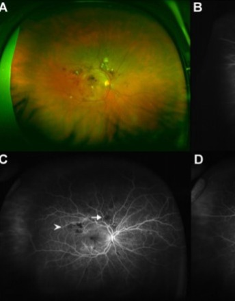
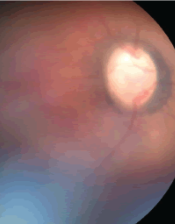
Congenital cavitary optic disc anomaly in Wolf-Hirschhorn Syndrome
Wolf-Hirschhorn Syndrome (WHS, OMIM 194190) is a rare congenital malformation syndrome caused by a partial deletion of the short arm (p) of chromosome 4. It is characterized by “Greek warrior helmet” facies, central nervous system disorders including seizures and structural defects, and intrauterine growth restriction, among numerous other systemic anomalies.
Evaluating refractive outcomes after pars plana vitrectomy and scleral fixated intraocular lens with Gore-Tex suture
It has been nearly 20 years since Girard first described the technique of placing a lens in the absence of capsular support [1]. Since that first report, numerous procedures have been described [2-9]. While the techniques have continued to evolve, little has been reported on refractive outcomes.
Visual field defect as an idiosyncratic reaction to topiramate
Topiramate is an increasingly popular medication used in the treatment of migraines, seizures, and other neurologic disorders. Its several mechanisms of action include enhancement of postsynaptic gamma-aminobutyric acid (GABA) receptor activity (an inhibitory neurotransmitter) and mild inhibition of carbonic anhydrase isoenzymes.
From observation to diagnosis: Implications of calcified sclero-choroidal choristomas in mosaic RASopathies
The identification of calcified sclero-choroidal choristomas (CaSCCs) in patients with mosaic RASopathies introduces a new dimension to understanding ocular manifestations in these genetic disorders.
An atypical case of compressive optic neuropathy and cranial nerve 6th palsy caused by a cholesterol granuloma
In this case report we present an unusual case of orbital cholesterol granuloma associated with compressive optic neuropathy and cranial nerve 6th palsy. Cholesterol granuloma results from a foreign body response to the presence of crystallized cholesterol. Cholesterol granuloma affecting the orbit are a rare presentation as they typically occur in the petrous apex of the temporal bone.
Ocular cystinosis – A review of disease, diagnosis, and future treatment options
Cystinosis is a rare autosomal recessive lysosomal storage disorder, characterised by the intra-lysosomal accumulation of cystine. Cystinosis results from a defect in the CTNS protein, a lysosomal transport protein for cystine. There are three subtypes of cystinosis: infantile nephropathic cystinosis, juvenile nephropathic cystinosis and ocular non-nephropathic cystinosis.